2023-02-28 エディンバラ大学
この研究結果は、Annals of Neurology誌に掲載されました。
<関連情報>
- https://www.ed.ac.uk/news/2023/breast-milk-boost-premature-baby-brain-development
- https://onlinelibrary.wiley.com/doi/10.1002/ana.26559
母乳への曝露は早産児の皮質成熟と関連する Breast Milk Exposure is Associated With Cortical Maturation in Preterm Infants
Gemma Sullivan, Kadi Vaher, MRes, Manuel Blesa , Paola Galdi, David Q. Stoye, Alan J. Quigley,Michael J. Thrippleton, John Norrie, Mark E. Bastin, James P. Boardman
Annals of Neurology Published: 22 November 2022
DOI:https://doi.org/10.1002/ana.26559

Abstract
Objective
Breast milk exposure is associated with improved neurocognitive outcomes following preterm birth but the neural substrates linking breast milk with outcome are uncertain. We tested the hypothesis that high versus low breast milk exposure in preterm infants results in cortical morphology that more closely resembles that of term-born infants.
Methods
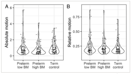
We studied 135 preterm (<32 weeks’ gestation) and 77 term infants. Feeding data were collected from birth until hospital discharge and brain magnetic resonance imaging (MRI) was performed at term-equivalent age. Cortical indices (volume, thickness, surface area, gyrification index, sulcal depth, and curvature) and diffusion parameters (fractional anisotropy [FA], mean diffusivity [MD], radial diffusivity [RD], axial diffusivity [AD], neurite density index [NDI], and orientation dispersion index [ODI]) were compared between preterm infants who received exclusive breast milk for <75% of inpatient days, preterm infants who received exclusive breast milk for ≥75% of inpatient days and term-born controls. To investigate a dose response effect, we performed linear regression using breast milk exposure quartile weighted by propensity scores.
Results
In preterm infants, high breast milk exposure was associated with reduced cortical gray matter volume (d = 0.47, 95% confidence interval [CI] = 0.14 to 0.94, p = 0.014), thickness (d = 0.42, 95% CI = 0.08 to 0.84, p = 0.039), and RD (d = 0.38, 95% CI = 0.002 to 0.77, p = 0.039), and increased FA (d = -0.38, 95% CI = -0.74 to -0.01, p = 0.037) after adjustment for age at MRI, which was similar to the cortical phenotype observed in term-born controls. Breast milk exposure quartile was associated with cortical volume (ß = -0.192, 95% CI = -0.342 to -0.042, p = 0.017), FA (ß = 0.223, 95% CI = 0.075 to 0.372, p = 0.007), and RD (ß = -0.225, 95% CI = -0.373 to -0.076, p = 0.007) following adjustment for age at birth, age at MRI, and weighted by propensity scores, suggesting a dose effect.
Interpretation
High breast milk exposure following preterm birth is associated with a cortical imaging phenotype that more closely resembles the brain morphology of term-born infants and effects appear to be dose-dependent. ANN NEUROL 2023;93:591–603


