
2024-05-30 カリフォルニア工科大学(Caltech)
")
Schematic showing location of cranial prosthetic and area of brain that can be imaged through this prosthetic.
<関連情報>
- https://www.caltech.edu/about/news/window-into-the-brain
- https://www.science.org/doi/10.1126/scitranslmed.adj3143
- https://www.nature.com/articles/s41593-023-01500-7
- https://www.cell.com/neuron/fulltext/S0896-6273(21)00151-3
音響的に透明な頭蓋窓を通したヒトの脳活動の機能的超音波イメージング Functional ultrasound imaging of human brain activity through an acoustically transparent cranial window
CLAIRE RABUT, SUMNER L. NORMAN, WHITNEY S. GRIGGS, JONATHAN J. RUSSIN, […], AND MIKHAIL G. SHAPIRO
Science Translational Medicine Published:29 May 2024
DOI:https://doi.org/10.1126/scitranslmed.adj3143
Editor’s summary
Imaging of human brain activity has the potential to unlock new ways to evaluate neurological function and disease. Despite substantial technological advances, the ability to sensitively and noninvasively record human brain activity in awake individuals has been difficult to achieve. Here, Rabut et al. report that a polymeric cranial window, implanted as part of skull reconstruction after traumatic brain injury in an adult individual, enabled the use of functional ultrasound imaging to measure brain activity in an adult human undertaking a task. This study provides proof of principle for the use of ultrasound imaging through transparent skull replacement materials to gain insight into human brain activity. —Molly Ogle
Abstract
Visualization of human brain activity is crucial for understanding normal and aberrant brain function. Currently available neural activity recording methods are highly invasive, have low sensitivity, and cannot be conducted outside of an operating room. Functional ultrasound imaging (fUSI) is an emerging technique that offers sensitive, large-scale, high-resolution neural imaging; however, fUSI cannot be performed through the adult human skull. Here, we used a polymeric skull replacement material to create an acoustic window compatible with fUSI to monitor adult human brain activity in a single individual. Using an in vitro cerebrovascular phantom to mimic brain vasculature and an in vivo rodent cranial defect model, first, we evaluated the fUSI signal intensity and signal-to-noise ratio through polymethyl methacrylate (PMMA) cranial implants of different thicknesses or a titanium mesh implant. We found that rat brain neural activity could be recorded with high sensitivity through a PMMA implant using a dedicated fUSI pulse sequence. We then designed a custom ultrasound-transparent cranial window implant for an adult patient undergoing reconstructive skull surgery after traumatic brain injury. We showed that fUSI could record brain activity in an awake human outside of the operating room. In a video game “connect the dots” task, we demonstrated mapping and decoding of task-modulated cortical activity in this individual. In a guitar-strumming task, we mapped additional task-specific cortical responses. Our proof-of-principle study shows that fUSI can be used as a high-resolution (200 μm) functional imaging modality for measuring adult human brain activity through an acoustically transparent cranial window.
閉ループ超音波ブレイン・マシン・インターフェースを用いた運動計画の解読 Decoding motor plans using a closed-loop ultrasonic brain–machine interface
Whitney S. Griggs,Sumner L. Norman,Thomas Deffieux,Florian Segura,Bruno-Félix Osmanski,Geeling Chau,Vasileios Christopoulos,Charles Liu,Mickael Tanter,Mikhail G. Shapiro & Richard A. Andersen
Nature Neuroscience Published:30 November 2023
DOI:https://doi.org/10.1038/s41593-023-01500-7
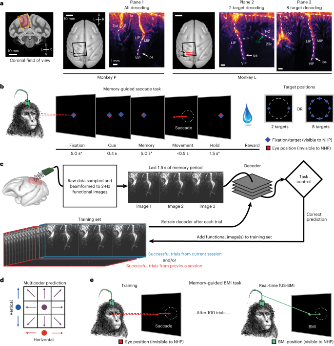
Abstract
Brain–machine interfaces (BMIs) enable people living with chronic paralysis to control computers, robots and more with nothing but thought. Existing BMIs have trade-offs across invasiveness, performance, spatial coverage and spatiotemporal resolution. Functional ultrasound (fUS) neuroimaging is an emerging technology that balances these attributes and may complement existing BMI recording technologies. In this study, we use fUS to demonstrate a successful implementation of a closed-loop ultrasonic BMI. We streamed fUS data from the posterior parietal cortex of two rhesus macaque monkeys while they performed eye and hand movements. After training, the monkeys controlled up to eight movement directions using the BMI. We also developed a method for pretraining the BMI using data from previous sessions. This enabled immediate control on subsequent days, even those that occurred months apart, without requiring extensive recalibration. These findings establish the feasibility of ultrasonic BMIs, paving the way for a new class of less-invasive (epidural) interfaces that generalize across extended time periods and promise to restore function to people with neurological impairments.
機能的超音波ニューロイメージングを用いた運動意図の単一試行デコーディング Single-trial decoding of movement intentions using functional ultrasound neuroimaging
Sumner L. Norman,David Maresca ,Vassilios N. Christopoulos ,Mickael Tanter,…,Mikhail G. Shapiro,Richard A. Andersen
Neuron Published:March 22, 2021
DOI:https://doi.org/10.1016/j.neuron.2021.03.003

Highlights
- Functional ultrasound (fUS) images motor planning activity in non-human primates
- fUS neuroimaging can predict single-trial movement timing, direction, and effector
- This is a critical step toward less-invasive and scalable brain-machine interfaces
Summary
New technologies are key to understanding the dynamic activity of neural circuits and systems in the brain. Here, we show that a minimally invasive approach based on ultrasound can be used to detect the neural correlates of movement planning, including directions and effectors. While non-human primates (NHPs) performed memory-guided movements, we used functional ultrasound (fUS) neuroimaging to record changes in cerebral blood volume with 100 μm resolution. We recorded from outside the dura above the posterior parietal cortex, a brain area important for spatial perception, multisensory integration, and movement planning. We then used fUS signals from the delay period before movement to decode the animals’ intended direction and effector. Single-trial decoding is a prerequisite to brain-machine interfaces, a key application that could benefit from this technology. These results are a critical step in the development of neuro-recording and brain interface tools that are less invasive, high resolution, and scalable.

")