2025-04-16 中国科学院(CAS)

<関連情報>
- https://english.cas.cn/newsroom/research_news/life/202504/t20250423_1041763.shtml
- https://direct.mit.edu/imag/article/doi/10.1162/imag_a_00561/128819/Attenuated-and-delayed-neural-activity-in-cortical
ヒト弱視における単眼処理と両眼相互作用の皮質微小回路における神経活動の減衰と遅延 Attenuated and delayed neural activity in cortical microcircuitry of monocular processing and binocular interactions in human amblyopia
Yue Wang,Chencan Qian,Yige Gao,Yulian Zhou,Xiaotong Zhang,Wen Wen,Peng Zhang
Imaging Neuroscience Published:April 11 2025
DOI:https://doi.org/10.1162/imag_a_00561

Abstract
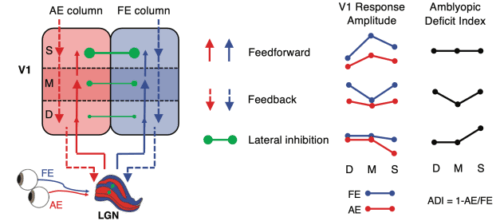
Disruption of retinal input early in life can lead to amblyopia, a condition characterized by reduced visual acuity after optical correction. While functional abnormalities in the early visual areas have been observed in amblyopia, mesoscale deficits in cortical microcircuitry across cortical depth remain unexplored in humans. Using a combination of submillimeter 7T fMRI and EEG frequency-tagging methods, we investigated neural deficits in monocular processing and binocular interactions in human adults with unilateral amblyopia. The results revealed attenuated and delayed monocular activity in the thalamic input layers of V1, followed by imbalanced binocular suppression and weakened binocular integration in the superficial layers. These disruptions further reduced visual signal strength and processing speed. Our findings pinpoint specific neural deficits in the cortical microcircuitry associated with human amblyopia, offering valuable insights into the mesoscale mechanisms of developmental plasticity and paving the way for more effective treatments for this visual disorder.

