新しいイメージング技術で、発達中の脳の循環パターンを解明 New imaging technique reveals circulation patterns in developing brain
2023-02-09 ワシントン大学セントルイス校
◆ワシントン大学医学部(セントルイス)の研究者らは、脳内の体液循環パターンを追跡する新しい技術を開発し、ネズミの実験で、正常な脳の発達と機能に重要な部位に脳液が流れていることを発見しました。さらに、子どもの認知障害と関連する水頭症の幼若ラットでは、循環に異常が見られることも発見されました。
◆この研究結果は、Nature Communications誌のオンライン版で公開されており、脳を満たしている液体(脳脊髄液)が、正常な脳の発達や神経発達障害において、十分に認識されていない役割を担っている可能性があることを示唆しています。
◆「脳脊髄液の力学の乱れは、水頭症やその他の発達性脳障害の子供に見られる脳の発達の変化の原因かもしれない」と、筆頭著者のジェニファー・ストラーレ、MD、脳神経外科、小児科、整形外科の准教授は述べている。小児神経外科医として、ストラーレはセントルイス小児病院で水頭症の子供たちを治療しています。「水頭症をはじめ、幼い子どもたちの神経疾患には、発達の遅れを伴うものがたくさんあります。これらの疾患の多くは、発達遅滞の根本的な原因がわかっていません。これらの症例の中には、脳脊髄液が循環している脳部位の機能に変化が生じている可能性があります。
◆成人の脳における脳脊髄液の排出をマッピングする多くの研究が行われています。しかし、脳脊髄液が脳そのものとどのように相互作用しているかはよく分かっていません。幼い子どもは、大人のような成熟した排液経路がまだ発達していないため、脳内の脳脊髄液の経路は年齢によって異なる可能性が高いのです。
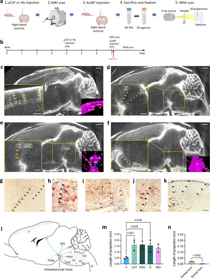
◆研究チームは、金ナノ粒子を用いたX線イメージング技術を開発し、脳の循環パターンを微細に可視化することに成功した。この方法を若いマウスとラットに適用したところ、脳脊髄液が主に脳の底部にある小さな流路を通って脳に入り込むことがわかった(この流路は成人では見られない)。さらに、脳脊髄液は脳の特定の機能部位に流れていることを発見した。
◆更なる実験が、水頭症が、明確なニューロン集団への脳脊髄液の流れを減少させることを示しました。Strahleたちは、一部の未熟児が罹患する水頭症の一種を研究した。早産で生まれた赤ちゃんは、出生前後に脳出血を起こしやすく、水頭症や発達の遅れにつながる可能性がある。ストラールらは、未熟児のプロセスを模倣したプロセスを若いラットに誘導した。3日後、脳の外表面から中央に脳脊髄液を運ぶ小さなチャネルが少なくなり、短くなり、24の神経細胞群のうち15への循環が著しく減少していたのである。
<関連情報>
- https://source.wustl.edu/2023/02/disrupted-flow-of-brain-fluid-may-underlie-neurodevelopmental-disorders/
- https://medicine.wustl.edu/news/disrupted-flow-of-brain-fluid-may-underlie-neurodevelopmental-disorders/
- https://www.nature.com/articles/s41467-023-36083-1
金ナノ粒子増強X線マイクロトモグラフィによるげっ歯類の脳内領域特異的脳脊髄液循環の解明 Gold nanoparticle-enhanced X-ray microtomography of the rodent reveals region-specific cerebrospinal fluid circulation in the brain
Shelei Pan,Peter H. Yang,Dakota DeFreitas,Sruthi Ramagiri,Peter O. Bayguinov,Carl D. Hacker,Abraham Z. Snyder,Jackson Wilborn,Hengbo Huang,Gretchen M. Koller,Dhvanii K. Raval,Grace L. Halupnik,Sanja Sviben,Samuel Achilefu,Rui Tang,Gabriel Haller,James D. Quirk,James A. J. Fitzpatrick,Prabagaran Esakky & Jennifer M. Strahle
Nature Communications Published:27 January 2023
DOI:https://doi.org/10.1038/s41467-023-36083-1

Abstract
Cerebrospinal fluid (CSF) is essential for the development and function of the central nervous system (CNS). However, the brain and its interstitium have largely been thought of as a single entity through which CSF circulates, and it is not known whether specific cell populations within the CNS preferentially interact with the CSF. Here, we develop a technique for CSF tracking, gold nanoparticle-enhanced X-ray microtomography, to achieve micrometer-scale resolution visualization of CSF circulation patterns during development. Using this method and subsequent histological analysis in rodents, we identify previously uncharacterized CSF pathways from the subarachnoid space (particularly the basal cisterns) that mediate CSF-parenchymal interactions involving 24 functional-anatomic cell groupings in the brain and spinal cord. CSF distribution to these areas is largely restricted to early development and is altered in posthemorrhagic hydrocephalus. Our study also presents particle size-dependent CSF circulation patterns through the CNS including interaction between neurons and small CSF tracers, but not large CSF tracers. These findings have implications for understanding the biological basis of normal brain development and the pathogenesis of a broad range of disease states, including hydrocephalus.


