2024-11-08 ミュンヘン大学(LMU)
<関連情報>
- https://www.lmu.de/en/newsroom/news-overview/news/microscopy-vertical-dna-in-motion.html
- https://www.nature.com/articles/s41592-024-02498-x
蛍光顕微鏡で垂直配列DNAを用いた1分子動的構造生物学 Single-molecule dynamic structural biology with vertically arranged DNA on a fluorescence microscope
Alan M. Szalai,Giovanni Ferrari,Lars Richter,Jakob Hartmann,Merve-Zeynep Kesici,Bosong Ji,Kush Coshic,Martin R. J. Dagleish,Annika Jaeger,Aleksei Aksimentiev,Ingrid Tessmer,Izabela Kamińska,Andrés M. Vera & Philip Tinnefeld
Nature Methods Published:08 November 2024
DOI:https://doi.org/10.1038/s41592-024-02498-x

Abstract
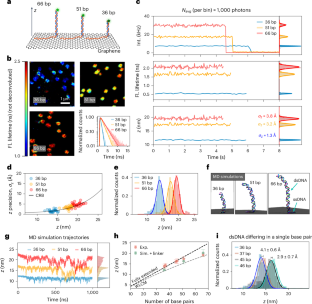
The intricate interplay between DNA and proteins is key for biological functions such as DNA replication, transcription and repair. Dynamic nanoscale observations of DNA structural features are necessary for understanding these interactions. Here we introduce graphene energy transfer with vertical nucleic acids (GETvNA), a method to investigate DNA–protein interactions that exploits the vertical orientation adopted by double-stranded DNA on graphene. This approach enables the dynamic study of DNA conformational changes via energy transfer from a probe dye to graphene, achieving spatial resolution down to the Ångström scale at subsecond temporal resolution. We measured DNA bending induced by adenine tracts, bulges, abasic sites and the binding of endonuclease IV. In addition, we observed the translocation of the O6-alkylguanine DNA alkyltransferase on DNA, reaching single base-pair resolution and detecting preferential binding to adenine tracts. This method promises widespread use for dynamical studies of nucleic acids and nucleic acid–protein interactions with resolution so far reserved for traditional structural biology techniques.


