MITとブラウン大学のチームは、磁気センサーを使って運動中の筋肉の長さを追跡することの正確さと安全性を実証し、切断手術を受けた人が義肢を容易に操作できるようになる可能性を示しました。 Team from MIT and Brown demonstrates the accuracy and safety of using magnetic sensors to track muscle length during movement, which could make it easier for people with amputations to control prosthetic limbs.
2022-10-31 ブラウン大学
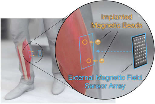
研究者らは、発表した2本の論文の中で、運動中の筋肉の長さを追跡できる磁石ベースのシステムの精度と安全性を実証している。この研究は、動物モデルを用いて行われ、この方法を用いることで、義肢装具を装着した人がより自然な手足の動きに近い形で義肢装具を操作できるようになる可能性を示している。
研究チームは、七面鳥が登るためのスロープと、飛び乗ったり降りたりするための箱からなる障害物コースを作成した。研究チームは、磁気センサーを使ってこれらの活動中の筋肉の動きを追跡し、システムが1ミリ秒未満で筋肉の長さを計算できることを発見した。
また、磁気センサよりも大型の装置を必要とするX線技術の一種である蛍光X線分析法という従来の手法で測定したデータと比較した。その結果、磁気マイクロメトリクスの測定値は、蛍光マイクロメトリクスの測定値と平均して1ミリメートル未満の差しか生じなかった。
室内で使用するX線装置と同等の筋長追跡機能を、より小型で持ち運び可能なパッケージで実現し、さらに、蛍光X線分析装置のように10秒間の連続測定に制限されることなく、連続的にデータを収集できる。
2つ目の論文では、インプラントの生体適合性に着目している。その結果、磁石は組織の瘢痕化や炎症などの有害な影響を及ぼさないことがわかった。また、磁石が七面鳥の歩行を変化させないことから、磁石が不快感を与えないことも明らかにした。
さらに、8カ月間の試験期間中、磁石は安定しており、3センチメートル以上離して埋め込めば、互いに移動しないことも確認された。
磁石は外部電源を必要としないので、筋肉に埋め込んだ後、患者の生涯を通じて磁場の強さを完全に維持することができる。
<関連情報>
- https://www.brown.edu/news/2022-10-31/magnetomicrometry
- https://www.frontiersin.org/articles/10.3389/fbioe.2022.1010275/full
- https://www.frontiersin.org/articles/10.3389/fbioe.2022.1010276/full
マグネトミクロメトリを用いた非結束型筋電図 Untethered muscle tracking using magnetomicrometry
Cameron R. Taylor, Seong Ho Yeon, William H. Clark, Ellen G. Clarrissimeaux, Mary Kate O’Donnell, Thomas J. Roberts and Hugh M. Herr
Frontiers in Bioengineering and Biotechnology Published:25 October 2022
DOI:https://doi.org/10.3389/fbioe.2022.1010275
")
Muscle tissue drives nearly all movement in the animal kingdom, providing power, mobility, and dexterity. Technologies for measuring muscle tissue motion, such as sonomicrometry, fluoromicrometry, and ultrasound, have significantly advanced our understanding of biomechanics. Yet, the field lacks the ability to monitor muscle tissue motion for animal behavior outside the lab. Towards addressing this issue, we previously introduced magnetomicrometry, a method that uses magnetic beads to wirelessly monitor muscle tissue length changes, and we validated magnetomicrometry via tightly-controlled in situ testing. In this study we validate the accuracy of magnetomicrometry against fluoromicrometry during untethered running in an in vivo turkey model. We demonstrate real-time muscle tissue length tracking of the freely-moving turkeys executing various motor activities, including ramp ascent and descent, vertical ascent and descent, and free roaming movement. Given the demonstrated capacity of magnetomicrometry to track muscle movement in untethered animals, we feel that this technique will enable new scientific explorations and an improved understanding of muscle function.
筋肉への磁気ビーズインプラントの臨床的有用性 Clinical viability of magnetic bead implants in muscle
Cameron R. Taylor, William H. Clark, Ellen G. Clarrissimeaux, Seong Ho Yeon, Matthew J. Carty, Stuart R. Lipsitz, Roderick T. Bronson, Thomas J. Roberts and Hugh M. Herr
Frontiers in Bioengineering and Biotechnology Published:25 October 2022
DOI:https://doi.org/10.3389/fbioe.2022.1010276
Human movement is accomplished through muscle contraction, yet there does not exist a portable system capable of monitoring muscle length changes in real time. To address this limitation, we previously introduced magnetomicrometry, a minimally-invasive tracking technique comprising two implanted magnetic beads in muscle and a magnetic field sensor array positioned on the body’s surface adjacent the implanted beads. The implant system comprises a pair of spherical magnetic beads, each with a first coating of nickel-copper-nickel and an outer coating of Parylene C. In parallel work, we demonstrate submillimeter accuracy of magnetic bead tracking for muscle contractions in an untethered freely-roaming avian model. Here, we address the clinical viability of magnetomicrometry. Using a specialized device to insert magnetic beads into muscle in avian and lagomorph models, we collect data to assess gait metrics, bead migration, and bead biocompatibility. For these animal models, we find no gait differences post-versus pre-implantation, and bead migration towards one another within muscle does not occur for initial bead separation distances greater than 3 cm. Further, using extensive biocompatibility testing, the implants are shown to be non-irritant, non-cytotoxic, non-allergenic, and non-irritating. Our cumulative results lend support for the viability of these magnetic bead implants for implantation in human muscle. We thus anticipate their imminent use in human-machine interfaces, such as in control of prostheses and exoskeletons and in closed-loop neuroprosthetics to aid recovery from neurological disorders.
")
")