2022-11-11 スイス連邦工科大学ローザンヌ校(EPFL)
顕微鏡で見ると、健康な細胞とそうでない細胞を区別するのは非常に難しい。科学者は、特定のタンパク質を標的とした染色や蛍光タグを用いて、細胞の種類を特定し、その状態を特徴づけ、薬剤や他の治療法の影響を研究している。この方法は医学に大きな影響を与えたが、一方で限界もある。1つは、細胞へのタグ付けは高価で時間がかかり、研究者の技量に強く依存することだ。その上、染色工程は研究対象の細胞に悪影響を与える可能性がある。
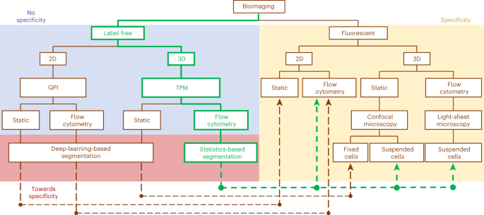
Nature Photonics誌に掲載された論文では、生きた細胞内の特定の領域を正確に識別できる無染色法を発表している。ホログラフィックイメージングとマイクロ流体工学をニューラルネットワークベースの信号処理と独自に組み合わせたこの研究は、循環腫瘍細胞検出のための液体生検や薬物検査のための高スループットアッセイへの道を開くものである。
ラーニング・トモグラフィーは、顕微鏡を使って研究対象の視覚的画像を作成するのではなく、ホログラフィック・イメージング法を用いて、顕微鏡の光ビームが細胞を構成する物質を通過する際に生じる位相遅れを明らかにする。
研究チームは、このプロセスをさまざまな角度で繰り返し、位相データをニューラルネットワークにかけることで、個々のボクセル(この手法で解像された3次元ボリューム)の屈折率の3Dマップを作成することができた。
屈折率の近いボクセルをグループ化する自己クラスタリング手法と機械学習ツールを用いることで、クラスタを分類可能な形状に組み立てることができた。
50~100ミクロンの流路に細胞を入れ、流路内の流速勾配で細胞を回転させ、固定ビームと検出器を用いて、細胞が流路に沿って回転する様子を観察することで、位相差を検出し、細胞の向きを推定し、学習型トモグラフィーを適用して3D屈折率マップを作成することができる。
<関連情報>
- https://actu.epfl.ch/news/researchers-open-door-to-stain-free-labeling-of-ce/
- https://www.nature.com/articles/s41566-022-01096-7#citeas
フローサイトメトリーにおけるトモグラフィ位相顕微鏡を用いた無染色での細胞核の同定
Stain-free identification of cell nuclei using tomographic phase microscopy in flow cytometry
Daniele Pirone,Joowon Lim,Francesco Merola,Lisa Miccio,Martina Mugnano,Vittorio Bianco,Flora Cimmino,Feliciano Visconte,Annalaura Montella,Mario Capasso,Achille Iolascon,Pasquale Memmolo,Demetri Psaltis & Pietro Ferraro
Nature Photonics Published:10 November 2022
DOI:https://doi.org/10.1038/s41566-022-01096-7

Abstract
Quantitative phase imaging has gained popularity in bioimaging because it can avoid the need for cell staining, which, in some cases, is difficult or impossible. However, as a result, quantitative phase imaging does not provide the labelling of various specific intracellular structures. Here we show a novel computational segmentation method based on statistical inference that makes it possible for quantitative phase imaging techniques to identify the cell nucleus. We demonstrate the approach with refractive index tomograms of stain-free cells reconstructed using tomographic phase microscopy in the flow cytometry mode. In particular, by means of numerical simulations and two cancer cell lines, we demonstrate that the nucleus can be accurately distinguished within the stain-free tomograms. We show that our experimental results are consistent with confocal fluorescence microscopy data and microfluidic cyto-fluorimeter outputs. This is a remarkable step towards directly extracting specific three-dimensional intracellular structures from the phase contrast data in a typical flow cytometry configuration.


