2023-01-16 オランダ・デルフト工科大学(TUDelft)
より優れた骨インプラント
そのアプローチの1つは、バイオマテリアルの物理的表面の手がかりに着目し、小さな柱などの異なる表面形状が、細胞に「私たちがさせたいこと」(この場合は骨組織の再生)をどのように指示するかを確認することである。「課題は、骨細胞になるための前駆細胞の接着と分化を支援すると同時に、細菌細胞の増殖を抑制し、インプラントに関連する感染を防ぐようなパターンを表面に作り出すことです。骨細胞が好むように、しかし細菌が好まないように表面を構造化するのです。これは非常に巧妙なアイデアで、私を魅了しました」とガトケサーは語っています。
コヒーシヨン・プロジェクト
Fratila-ApachiteiとGhatkesar(PME)は、サブマイクロメートル領域の特徴をもつ3Dプリント柱が、生きた前駆細胞の接着と力学にどのような影響を与えるかを単一細胞レベルで調べるため、Cohesion補助金に応募し、これを獲得した。ポスドク研究員のLivia Angeloniと他の共同研究者と共に、流体力顕微鏡に基づいた新しい方法を開発し、適用しました。
半ミクロメートルが重要
“我々の方法を使うことで、骨形成分化とマトリックス鉱化に関する潜在的な生物物理学的マーカー(例えば、細胞接着強度)を早期に明らかにすることができ、0.5マイクロメートルの柱の高さのわずかな違いが、細胞の行動を変えるのに十分であることを示しています。” この方法と得られた結果は、整形外科用生体材料の物理的表面キューをより合理的に設計することを可能にします。彼らの結果は、Small誌に掲載され、本誌の表紙内側に掲載されています。
<関連情報>
- https://www.tudelft.nl/en/2023/3me/news/new-methods-to-investigate-the-interface-between-biomaterials-and-cells-to-help-regenerate-body-tissues
- https://onlinelibrary.wiley.com/doi/10.1002/smll.202204662
流体力顕微鏡と原子間力顕微鏡による3Dプリントされたサブミクロンパターンと前骨芽細胞の相互作用に関する新たな知見 Fluidic Force Microscopy and Atomic Force Microscopy Unveil New Insights into the Interactions of Preosteoblasts with 3D-Printed Submicron Patterns
Livia Angeloni, Bogdan Popa, Mahdiyeh Nouri-Goushki, Michelle Minneboo, Amir A. Zadpoor, Murali K. Ghatkesar, Lidy E. Fratila-Apachitei
Small Published: 14 November 2022
DOI:https://doi.org/10.1002/smll.202204662
Abstract
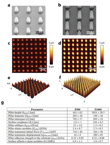
Physical patterns represent potential surface cues for promoting osteogenic differentiation of stem cells and improving osseointegration of orthopedic implants. Understanding the early cell–surface interactions and their effects on late cellular functions is essential for a rational design of such topographies, yet still elusive. In this work, fluidic force microscopy (FluidFM) and atomic force microscopy (AFM) combined with optical and electron microscopy are used to quantitatively investigate the interaction of preosteoblasts with 3D-printed patterns after 4 and 24 h of culture. The patterns consist of pillars with the same diameter (200 nm) and interspace (700 nm) but distinct heights (500 and 1000 nm) and osteogenic properties. FluidFM reveals a higher cell adhesion strength after 24 h of culture on the taller pillars (32 ± 7 kPa versus 21.5 ± 12.5 kPa). This is associated with attachment of cells partly on the sidewalls of these pillars, thus requiring larger normal forces for detachment. Furthermore, the higher resistance to shear forces observed for these cells indicates an enhanced anchorage and can be related to the persistence and stability of lamellipodia. The study explains the differential cell adhesion behavior induced by different pillar heights, enabling advancements in the rational design of osteogenic patterns.
")
")