2023-09-11 ワシントン大学セントルイス校

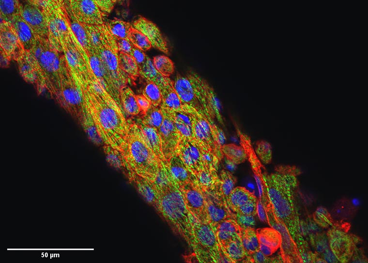
This image shows the microtissues from Nate Huebsch’s lab that are stained for cell nuclei (blue), sarcomeric alpha-actinic (red) and Myosin Binding Protein C (MYBPC3; green). Mutations in MYBPC3 cause hypertrophic cardiomyopathy. These sarcomere structures are disrupted by the chemical blebbistatin. (Image: Huebsch lab)
◆この研究は、心臓の電気伝導が機械的収縮とどのように関連しているかを直接的かつ非侵襲的に研究するためのツールを提供し、心疾患の理解に貢献します。疾患の中には、肥大型心筋症などがあり、この新しいアプローチは治療法の開発や治療効果の研究にも役立つ可能性があります。
<関連情報>
- https://engineering.wustl.edu/news/2023/Virtual-drug-quiets-noise-in-images-of-heart-tissue.html
- https://www.pnas.org/doi/10.1073/pnas.2212949120
- https://zenodo.org/record/7897265
仮想ブレッビスタチン:心筋細胞の光マッピングにおけるモーションアーチファクト除去のための堅牢かつ迅速なソフトウェアアプローチ Virtual blebbistatin: A robust and rapid software approach to motion artifact removal in optical mapping of cardiomyocytes
Louis G. Woodhams, Jingxuan Guo, David Schuftan, John J. Boyle, Kenneth M. Pryse, Elliot L. Elson, Nathaniel Huebsch, and Guy M. Genin
Proceedings of the National Academy of Sciences Published:September 11, 2023
DOI:https://doi.org/10.1073/pnas.2212949120
Significance
With advancements in stem cell technology, in vitro models using iPSC (induced pluripotent stem cells)-derived cardiomyocytes (iPSC-CM) and engineered heart tissues (EHT) can serve as powerful tools for disease modeling and drug screening. Optical methods can enable high throughput monitoring of tissue electrophysiology, but signals from voltage and calcium-sensitive dyes are susceptible to distortion by cardiac motion. Pharmacologic paralysis of tissues poses a fundamental barrier to study excitation–contraction coupling by disturbing contractility. Virtual removal of motion artifact and reconstruction of cardiac excitation and contraction coupling with lower computational cost and high accuracy will provide great opportunities to unveil mechanisms of acquired and inherited cardiac diseases and test potential drug therapies.
Abstract
Fluorescent reporters of cardiac electrophysiology provide valuable information on heart cell and tissue function. However, motion artifacts caused by cardiac muscle contraction interfere with accurate measurement of fluorescence signals. Although drugs such as blebbistatin can be applied to stop cardiac tissue from contracting by uncoupling calcium-contraction, their usage prevents the study of excitation–contraction coupling and, as we show, impacts cellular structure. We therefore developed a robust method to remove motion computationally from images of contracting cardiac muscle and to map fluorescent reporters of cardiac electrophysiological activity onto images of undeformed tissue. When validated on cardiomyocytes derived from human induced pluripotent stem cells (iPSCs), in both monolayers and engineered tissues, the method enabled efficient and robust reduction of motion artifact. As with pharmacologic approaches using blebbistatin for motion removal, our algorithm improved the accuracy of optical mapping, as demonstrated by spatial maps of calcium transient decay. However, unlike pharmacologic motion removal, our computational approach allowed direct analysis of calcium-contraction coupling. Results revealed calcium-contraction coupling to be more uniform across cells within engineered tissues than across cells in monolayer culture. The algorithm shows promise as a robust and accurate tool for optical mapping studies of excitation–contraction coupling in heart tissue.
バーチャルブレブスタチン画像安定化ソフトウェア Virtual Blebbistatin image stabilization software
Woodhams, Louis G.; Guo, Jingxuan; Schuftan, David; Boyle, John J.; Pryse, Kenneth M.; Elson, Elliot L.; Huebsch, Nathaniel; Genin, Guy M.
Zenodo Published:May 4, 2023
DOI:https://doi.org/10.5281/zenodo.7897265
This software takes a video or series of images, divides the initial frame into regions, maps the deformations of all regions onto subsequent frames, and stabilizes motion in frames by mapping pixel values in subsequent locations back their undeformed location.
For more information see the README file (included here) and the associated publication, Woodhams et al. (2023), Virtual Blebbistatin: A Robust and Rapid Software Approach to Motion Artifact Removal in Optical Mapping of Cardiomyocytes, Proceedings of the National Academy of Sciences, describing the application of Virtual Blebbistatin software.

