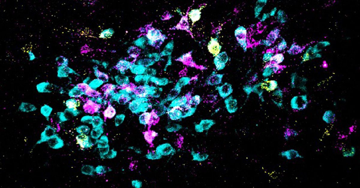
2024-03-14 カリフォルニア大学サンディエゴ校(UCSD)
<関連情報>
- https://today.ucsd.edu/story/new-imaging-tool-advances-study-of-lipid-biology
- https://www.nature.com/articles/s41467-024-45576-6
多分子ハイパースペクトルPRM-SRS顕微鏡法 Multi-molecular hyperspectral PRM-SRS microscopy
Wenxu Zhang,Yajuan Li,Anthony A. Fung,Zhi Li,Hongje Jang,Honghao Zha,Xiaoping Chen,Fangyuan Gao,Jane Y. Wu,Huaxin Sheng,Junjie Yao,Dorota Skowronska-Krawczyk,Sanjay Jain &Lingyan Shi
Nature Communications Published:21 February 2024
DOI:https://doi.org/10.1038/s41467-024-45576-6

Abstract
Lipids play crucial roles in many biological processes. Mapping spatial distributions and examining the metabolic dynamics of different lipid subtypes in cells and tissues are critical to better understanding their roles in aging and diseases. Commonly used imaging methods (such as mass spectrometry-based, fluorescence labeling, conventional optical imaging) can disrupt the native environment of cells/tissues, have limited spatial or spectral resolution, or cannot distinguish different lipid subtypes. Here we present a hyperspectral imaging platform that integrates a Penalized Reference Matching algorithm with Stimulated Raman Scattering (PRM-SRS) microscopy. Using this platform, we visualize and identify high density lipoprotein particles in human kidney, a high cholesterol to phosphatidylethanolamine ratio inside granule cells of mouse hippocampus, and subcellular distributions of sphingosine and cardiolipin in human brain. Our PRM-SRS displays unique advantages of enhanced chemical specificity, subcellular resolution, and fast data processing in distinguishing lipid subtypes in different organs and species.


