2023-09-21 スイス連邦工科大学ローザンヌ校(EPFL)
◆この新しいクライオEM方法は、タンパク質の機能を観察する重要な手段であり、将来的にはタンパク質の機能に関する理解を革新的に進化させる可能性があります。
<関連情報>
- https://actu.epfl.ch/news/new-imaging-technique-sees-virus-move-in-unprecede/
- https://actu.epfl.ch/news/new-imaging-technique-sees-virus-move-in-unprecede/
マイクロ秒時間分解低温電子顕微鏡で明らかになったウイルスの高速ダイナミクス Fast viral dynamics revealed by microsecond time-resolved cryo-EM
Oliver F. Harder,Sarah V. Barrass,Marcel Drabbels & Ulrich J. Lorenz
Nature Communications Published:13 September 2023
DOI:https://doi.org/10.1038/s41467-023-41444-x

Abstract
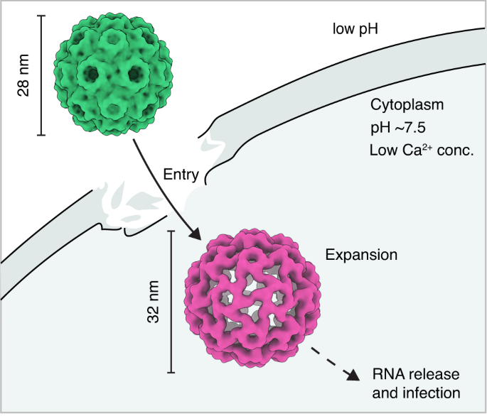
Observing proteins as they perform their tasks has largely remained elusive, which has left our understanding of protein function fundamentally incomplete. To enable such observations, we have recently proposed a technique that improves the time resolution of cryo-electron microscopy (cryo-EM) to microseconds. Here, we demonstrate that microsecond time-resolved cryo-EM enables observations of fast protein dynamics. We use our approach to elucidate the mechanics of the capsid of cowpea chlorotic mottle virus (CCMV), whose large-amplitude motions play a crucial role in the viral life cycle. We observe that a pH jump causes the extended configuration of the capsid to contract on the microsecond timescale. While this is a concerted process, the motions of the capsid proteins involve different timescales, leading to a curved reaction path. It is difficult to conceive how such a detailed picture of the dynamics could have been obtained with any other method, which highlights the potential of our technique. Crucially, our experiments pave the way for microsecond time-resolved cryo-EM to be applied to a broad range of protein dynamics that previously could not have been observed. This promises to fundamentally advance our understanding of protein function.


