2024-10-01 コロンビア大学

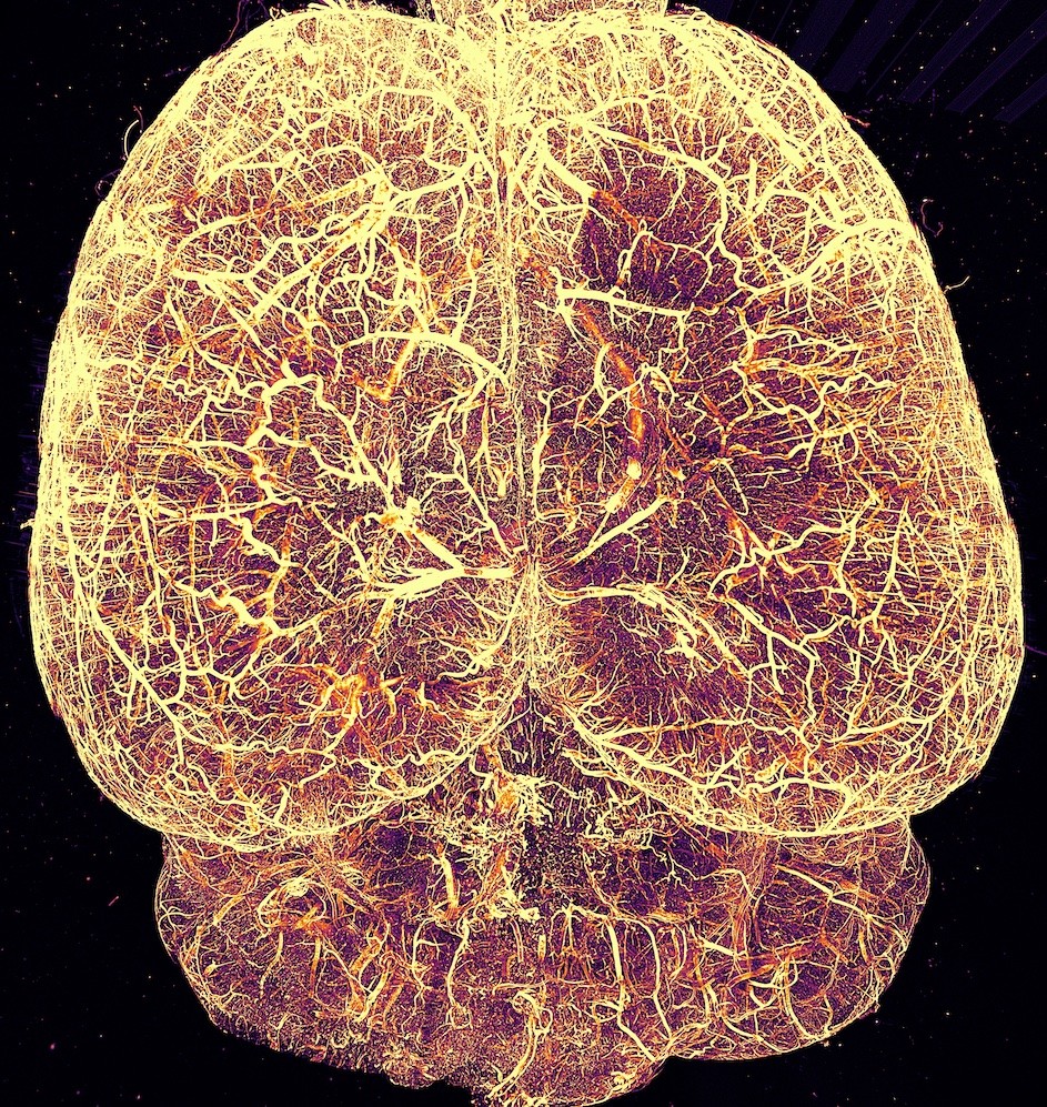
The entire vasculature of a mouse brain captured by Tomer’s new low-cost microscopy tool.
<関連情報>
- https://news.columbia.edu/news/developing-new-technologies-investigate-and-heal-brain
- https://www.nature.com/articles/s44222-024-00249-3
低コストで高性能なライトシート顕微鏡
Low-cost, high-performance light-sheet microscopy
Christine-Maria Horejs
Nature Reviews Bioengineering Published:17 September 2024
DOI:https://doi.org/10.1038/s44222-024-00249-3
Light-sheet fluorescence microscopy (LSFM) uses a thin sheet of light to excite only fluorophores within the focal volume, thereby enabling optical sectioning of a sample with minimal phototoxicity. Such selective illumination allows the long-term and quantitative mapping of molecules, cells and microenvironments within large, centimeter-scale samples made transparent through chemical tissue-clearing methods. Therefore, LSFM is particularly useful for the imaging and mapping of 3D biological objects — from organelles to organoids and organs. However, owing to its complex architecture, LSFM remains expensive and not easily scalable and is therefore not yet widely available.
The basic principle of LSFM involves illuminating a thin plane of the sample with a thin sheet of light and rapidly capturing images using a wide-field detection system positioned at a right angle. This setup considerably reduces the energy load during imaging and allows imaging speeds that are orders of magnitude faster than conventional point-scanning methods, such as confocal microscopy.


