2025-06-09 名古屋大学

<関連情報>
- https://www.nagoya-u.ac.jp/researchinfo/result/2025/06/post-833.html
- https://www.nagoya-u.ac.jp/researchinfo/result/upload_images/20250609_coi.pdf
- https://www.science.org/doi/10.1126/sciadv.adt6683
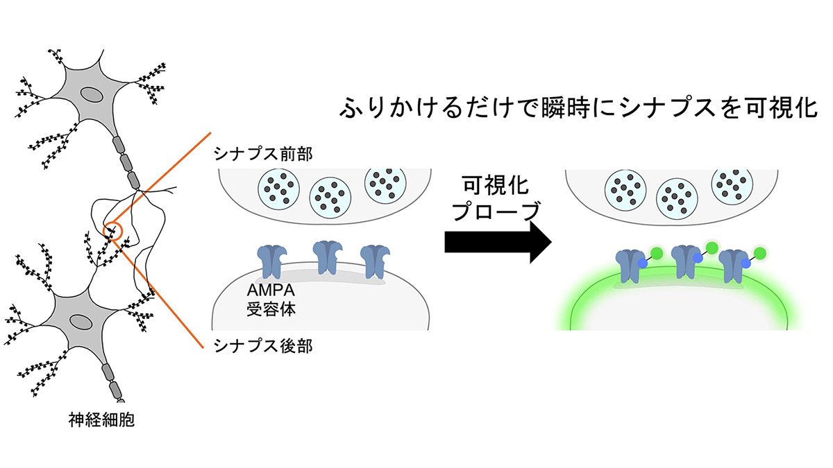
迅速かつ可逆的な蛍光プローブにより、シナプス可塑性におけるAMPA受容体の反復スナップショットイメージングが可能になる Rapid and reversible fluorescent probe enables repeated snapshot imaging of AMPA receptors during synaptic plasticity
Kyohei Soga, Takaaki Fujiwara, Mayu Nakagawa, Akihiro Shibata, […] , and Shigeki Kiyonaka
Science Advances Published:6 Jun 2025
DOI:https://doi.org/10.1126/sciadv.adt6683
Abstract
The subcellular localization of neurotransmitter receptors is strictly regulated in neurons. Changes in the trafficking of α-amino-3-hydroxy-5-methyl-4-isoxazolepropionic acid (AMPA)–type glutamate receptors (AMPARs) play an essential role in synaptic plasticity, which is the cellular basis of learning and memory. To explore receptor trafficking, genetically encoded approaches (e.g., the fusion of fluorescent proteins to receptors) are often used. However, concerns remain that genetic approaches cannot fully reproduce the receptor functions that are inherent to neurons. Herein, we report on PFQX1(AF488), a fluorescent probe for the visualization of cell-surface AMPARs without any genetic manipulation to neurons. The rapid and reversible staining features of this probe enabled snapshot imaging, which showed the accumulation of native AMPARs in dendritic spines during synaptic plasticity. Moreover, the mechanism of this synaptic accumulation, for which genetically encoded approaches have given controversial results, was revealed by integrating two chemical methods: PFQX1(AF488) and covalent chemical labeling.
