2025-06-12 東京農工大学

<関連情報>
- https://www.tuat.ac.jp/outline/disclosure/pressrelease/2025/20250612_01.html
- https://www.tuat.ac.jp/documents/tuat/outline/disclosure/pressrelease/2025/20250612_01.pdf
- https://onlinelibrary.wiley.com/doi/10.1002/smll.202503749
時空間細胞間コミュニケーションを可視化するサイトトランスデューサー Cytotransducers for Visualization of Spatiotemporal Intercellular Communication
Niko Kimura, Yoko Yamanishi, Shinya Sakuma
Small Published: 09 June 2025
DOI:https://doi.org/10.1002/smll.202503749
Abstract
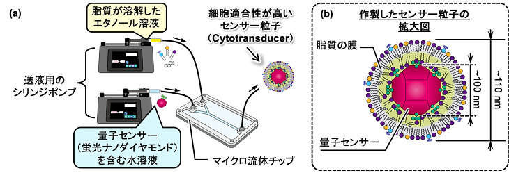
Visualization of intercellular communication provides a means for understanding and leveraging complicated multicellular organizations. Various nanometer-sized artifacts (nano-artifacts) can be employed as small tracers for visualization, achieving spatially and temporally controlled intervention of these small tracers in the interactions remains challenging due to their diffusion-based transportation. To achieve spatiotemporally controlled visualization, cytotransducers are conceptualized that convert veiled intra/intercellular information into visible information. A 110-mm cytotransducer is presented, composed of a lipid layer and a fluorescent nanodiamond. The designed lipid layer allows effective uptake of the cytotransducer by neutrophils, and the fluorescent nanodiamond is labeled with the pH-responsive chemical fluorescein isothiocyanate to visualize the intracellular local pH at the single-cell level. Utilizing cytotransducers, cross-scale visualization of intercellular communication between neutrophils and three types of cells: macrophages, aged cells, and mesenchymal stem cells is demonstrated. Intracellular pH maps are obtained before and during the interactions, achieving spatiotemporally controlled visualization of intercellular communication based on the localized release of cytotransducers by programmed cell death (NETosis). A well-designed cytotransducer allows the simultaneous visualization of intra/intercellular information under spatiotemporally controlled conditions.


