2022-04-05 コロンビア大学
・コロンビア大学のElizabeth Hillman博士が率いる研究チームは、Swept Confocally Aligned Planar Excitation Microscopy(SCAPE)と呼ばれるイメージングシステムを開発している。SCAPEは、すでにFDAが医療用システムとして承認しているレベルのレーザー光を使用します。組織全体にレーザーを照射し、組織内の特定の分子を発光させる。この光はレンズで集められ、高感度センサーに集光され、3D画像を形成する。
・研究チームはこれまでに、SCAPEが生体組織を迅速に画像化できることを実証している。今回の研究では、MediSCAPEと呼ばれるこの顕微鏡を、臨床の場で使用するための試験を行った。研究チームは、ベンチトップ型と手術室で使用するためのコンパクトなシステムの2つのプロトタイプを作成した。
・本研究結果は、2022年3月28日付のNature Biomedical Engineeringに掲載されました。

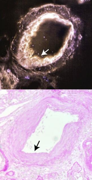
ヒト腎臓組織における動脈壁上のプラーク(矢印)。上段はMediSCAPEの画像、下段は従来の組織学的手法による同等の画像。 Patel et al., Nature Biomedical Engineering
<関連情報>
- https://www.nih.gov/news-events/nih-research-matters/system-reveals-3d-details-living-tissues
- https://pubmed.ncbi.nlm.nih.gov/35347275/
高速ライトシート顕微鏡による生体組織の体積組織像のin-situ取得 High-speed light-sheet microscopy for the in-situ acquisition of volumetric histological images of living tissue
Kripa B Patel, Wenxuan Liang, Malte J Casper, Venkatakaushik Voleti , Wenze Li , Alexis J Yagielski , Hanzhi T Zhao, Citlali Perez Campos, Grace Sooyeon Lee, Joyce M Liu, Elizabeth Philipone, Angela J Yoon, Kenneth P Olive, Shana M Coley, Elizabeth M C Hillman
Nature Biomedical Engineering Published:March 28, 2022
DOI: 10.1038/s41551-022-00849-7
Abstract
Histological examinations typically require the excision of tissue, followed by its fixation, slicing, staining, mounting and imaging, with timeframes ranging from minutes to days. This process may remove functional tissue, may miss abnormalities through under-sampling, prevents rapid decision-making, and increases costs. Here, we report the feasibility of microscopes based on swept confocally aligned planar excitation technology for the volumetric histological imaging of intact living tissue in real time. The systems’ single-objective, light-sheet geometry and 3D imaging speeds enable roving image acquisition, which combined with 3D stitching permits the contiguous analysis of large tissue areas, as well as the dynamic assessment of tissue perfusion and function. Implemented in benchtop and miniaturized form factors, the microscopes also have high sensitivity, even for weak intrinsic fluorescence, allowing for the label-free imaging of diagnostically relevant histoarchitectural structures, as we show for pancreatic disease in living mice, for chronic kidney disease in fresh human kidney tissues, and for oral mucosa in a healthy volunteer. Miniaturized high-speed light-sheet microscopes for in-situ volumetric histological imaging may facilitate the point-of-care detection of diverse cellular-level biomarkers.

