2022-09-11 スイス連邦工科大学ローザンヌ校(EPFL)
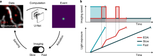
EPFLの生物物理学者たちは、人工ニューラルネットワークを利用して、試料へのストレスを抑えながら生物現象を詳細に撮影するための顕微鏡制御を自動化する方法を発見した。この技術は、バクテリアの細胞分裂にも、ミトコンドリアの分裂にも有効である。
研究チームは、ミトコンドリアの収縮(分裂につながるミトコンドリアの形状の変化)に注目するようニューラルネットワークを訓練し、分裂部位で豊富になることが知られているタンパク質の観測と組み合わせることで、細菌よりも難しいミトコンドリアの分裂を検出する方法を初めて解明した。
収縮とタンパク質の量がともに多い場合、顕微鏡は高速度撮影に切り替わり、分裂現象の詳細な画像を多数撮影することができる。収縮とタンパク質の量が少ないときは、低速度撮影に切り替えて、試料に過剰な光が当たらないようにする。
このインテリジェント蛍光顕微鏡を用いると、標準的な高速イメージングと比較して、より長い時間サンプルを観察することができることを示した。また、標準的な低速度撮影と比較して、サンプルに負荷がかかっているにもかかわらず、より意味のあるデータを取得することができた。
<関連情報>
- https://actu.epfl.ch/news/intelligent-microscopes-for-detecting-rare-biologi/
- https://www.nature.com/articles/s41592-022-01589-x
イベントドリブンアクイジションによるコンテンツエンリッチ顕微鏡の実現 Event-driven acquisition for content-enriched microscopy
Dora Mahecic,Willi L. Stepp,Chen Zhang,Juliette Griffié,Martin Weigert & Suliana Manley
Nature Methods Published:08 September 2022
DOI:https://doi.org/10.1038/s41592-022-01589-x

Abstract
A common goal of fluorescence microscopy is to collect data on specific biological events. Yet, the event-specific content that can be collected from a sample is limited, especially for rare or stochastic processes. This is due in part to photobleaching and phototoxicity, which constrain imaging speed and duration. We developed an event-driven acquisition framework, in which neural-network-based recognition of specific biological events triggers real-time control in an instant structured illumination microscope. Our setup adapts acquisitions on-the-fly by switching between a slow imaging rate while detecting the onset of events, and a fast imaging rate during their progression. Thus, we capture mitochondrial and bacterial divisions at imaging rates that match their dynamic timescales, while extending overall imaging durations. Because event-driven acquisition allows the microscope to respond specifically to complex biological events, it acquires data enriched in relevant content.


