照明環境下で大脳皮質全層の神経細胞活動を記録する小型装置を開発 Miniature device enables scientist to record nerve cell activity in all cortical layers in lit environments
2022-12-05 マックス・プランク研究所
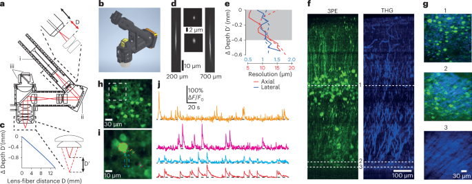
この研究では、2グラムの小型3光子励起顕微鏡が開発され、数々の初めての試みがなされた。動物の行動を妨げることなく、大脳皮質全層の神経活動を単一細胞レベルで画像化することが初めて可能になった。また、モジュール設計により、神経細胞の体細胞や樹状突起からの機能記録を可能にする高解像度構成となっている。また、検出器システムの改良により、完全な照明環境下でも使用できることも大きな特徴である。
研究チームは、この新しい小型3光子顕微鏡の視野と安定性を確認するため、マウスが自由にアリーナを探索している間に、皮質深部の第4層(L4)と第6層(L6)から画像を撮影した。その結果、L4とL6の神経細胞は、環境光によって異なる変調を受けることが判明した。顕微鏡は簡単に同じ位置に再設置できるため、同じ神経細胞集団を数日間に渡って追跡撮影することも可能である。これにより、例えば動物が学習している間の脳活動の変化をモニターできる可能性が出てきた。
<関連情報>
- https://www.mpg.de/19569467/1128-csar-neuroscientists-illuminate-how-brain-cells-deep-in-the-cortex-operate-in-freely-moving-mice-917443-x
- https://www.nature.com/articles/s41592-022-01688-9
自由に動くマウスの視覚野の全層をイメージングするための3光子ヘッドマウント顕微鏡 A three-photon head-mounted microscope for imaging all layers of visual cortex in freely moving mice
Alexandr Klioutchnikov,Damian J. Wallace,Juergen Sawinski,Kay-Michael Voit,Yvonne Groemping & Jason N. D. Kerr
Nature Methods Published:28 November 2022
DOI:https://doi.org/10.1038/s41592-022-01688-9

Abstract
Advances in head-mounted microscopes have enabled imaging of neuronal activity using genetic tools in freely moving mice but these microscopes are restricted to recording in minimally lit arenas and imaging upper cortical layers. Here we built a 2-g, three-photon excitation-based microscope, containing a z-drive that enabled access to all cortical layers while mice freely behaved in a fully lit environment. The microscope had on-board photon detectors, robust to environmental light, and the arena lighting was timed to the end of each line-scan, enabling functional imaging of activity from cortical layer 4 and layer 6 neurons expressing jGCaMP7f in mice roaming a fully lit or dark arena. By comparing the neuronal activity measured from populations in these layers we show that activity in cortical layer 4 and layer 6 is differentially modulated by lit and dark conditions during free exploration.


