胚細胞が力学的環境を感知して集合的に組織を形成する仕組みを解明 Researchers uncover how embryonic cells sense their mechanical environment to collectively form tissues
2022-12-28 カリフォルニア大学サンタバーバラ校(UCSB)
このような集団的な力学的感知は、細胞が分裂や移動、あるいは分化(幹細胞が特定の機能を果たすより特殊な細胞に変化する過程)を行うかどうかといった重要な決定を下すのに役立つ。数年前、合成基板上に置かれた幹細胞が、機械的な手がかりを頼りに意思決定を行うことが明らかになり、一つの大きな手がかりが得られた。骨に近い硬さの表面上の細胞は骨芽細胞(骨の細胞)になり、脳組織に近い硬さの表面上の細胞は神経細胞になったのです。この発見は、組織工学の分野を大きく前進させた。研究者たちはこの機械的な合図を使って合成足場を作り、幹細胞を望ましい結果に成長させるように誘導したのである。この足場は、現在ではさまざまな生物医学的用途に用いられている。
研究チームは、カンパス研究室で開発された独自のツールを用いて、胚の内部における細胞の本来の力学的環境を調べ、細胞が何になるかを決める際に知覚する物理量を明らかにすることに成功した。
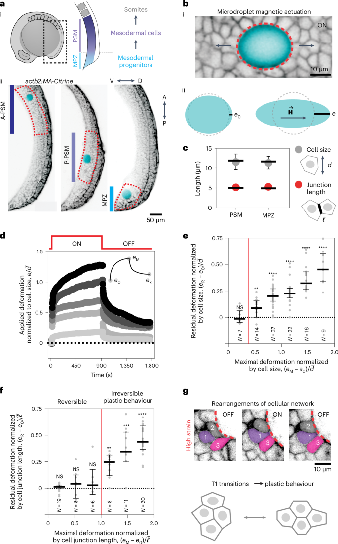
カンパス教授とその研究グループは、以前の論文(外部リンク)で、細胞が互いにくっついたり引っ張られたりしながら固まり、石鹸の泡やビールの泡に似た活発な泡の状態であることを発見している。カンパス教授とその研究チームは、細胞が機械的に探っているのは、個々の細胞やその中の構造体の硬さではなく、この「生きた泡」の集合状態、すなわち泡の硬さや集合体の閉じ込め具合であることを突き止めた。
この研究で得られた知見は、組織工学にとっても重要な意味を持つかもしれない。広く使われている合成ポリマーやゲルの足場とは対照的に、胚組織の泡状の特性を模倣した潜在的な材料があれば、研究者はより頑健で高度な合成組織、臓器、インプラントを、目的の機能に適した形状と力学的特性をもって、実験室で作り出すことができるようになるかもしれない。
<関連情報>
- https://www.news.ucsb.edu/2022/020798/stem-cells-sense-touch
- https://www.nature.com/articles/s41563-022-01433-9
ゼブラフィッシュ中胚葉前葉分化過程における生体内細胞からみた細胞微小環境のメカニクス Mechanics of the cellular microenvironment as probed by cells in vivo during zebrafish presomitic mesoderm differentiation
Alessandro Mongera,Marie Pochitaloff,Hannah J. Gustafson,Georgina A. Stooke-Vaughan,Payam Rowghanian,Sangwoo Kim & Otger Campàs
Nature Materials Published:28 December 2022
DOI:https://doi.org/10.1038/s41563-022-01433-9

Abstract
Tissue morphogenesis, homoeostasis and repair require cells to constantly monitor their three-dimensional microenvironment and adapt their behaviours in response to local biochemical and mechanical cues. Yet the mechanical parameters of the cellular microenvironment probed by cells in vivo remain unclear. Here, we report the mechanics of the cellular microenvironment that cells probe in vivo and in situ during zebrafish presomitic mesoderm differentiation. By quantifying both endogenous cell-generated strains and tissue mechanics, we show that individual cells probe the stiffness associated with deformations of the supracellular, foam-like tissue architecture. Stress relaxation leads to a perceived microenvironment stiffness that decreases over time, with cells probing the softest regime. We find that most mechanical parameters, including those probed by cells, vary along the anteroposterior axis as mesodermal progenitors differentiate. These findings expand our understanding of in vivo mechanosensation and might aid the design of advanced scaffolds for tissue engineering applications.


