2024-01-31 マサチューセッツ工科大学(MIT)

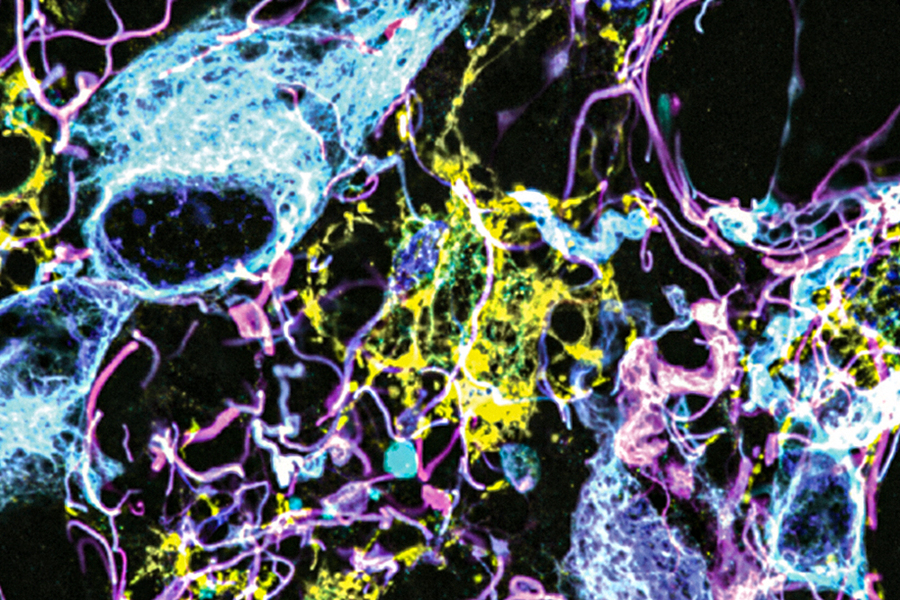
Using a novel microscopy technique, MIT and Harvard Medical School researchers have imaged human brain tissue in greater detail than ever before. In this image of a low-grade glioma, light blue and yellow represent different proteins associated with tumors. Pink indicates a protein used as a marker for astrocytes, and dark blue shows the location of cell nuclei.
Image: Courtesy of the researchers
◆この技術が将来的には腫瘍の診断、より正確な予後の算出、医師が治療法を選択するのに役立つことを期待しています。拡張顕微鏡法を使用して、保存されたヒト脳組織サンプルを拡大し、タンパク質のラベリングを行い、脳腫瘍や正常な脳組織の詳細な分析を実施。これにより、細胞間の相互作用や構造を従来のツールでは見えなかった範囲で理解できるようになり、脳腫瘍の悪性度をより正確に評価できる可能性があります。
<関連情報>
- https://news.mit.edu/2024/imaging-method-reveals-new-cells-structures-human-brain-tissue-0131
- https://www.science.org/doi/10.1126/scitranslmed.abo0049
ヒト脳標本中のナノ構造と細胞の免疫染色が、膨張を介したタンパク質の混雑緩和によって改善された Improved immunostaining of nanostructures and cells in human brain specimens through expansion-mediated protein decrowding
PABLO A. VALDES , CHIH-CHIEH (JAY) YU , JENNA ARONSON , DEBARATI GHOSH , […], AND EDWARD S. BOYDEN
Science Translational Medicine Published:31 Jan 2024
DOI:https://doi.org/10.1126/scitranslmed.abo0049
Editor’s summary
Expansion microscopy allows to image structures sized below the diffraction limit of light by physical expansion of biological specimens before imaging. Here, Valdes and colleagues developed decrowding expansion pathology (dExPath), a variant of expansion microscopy, that improved antibody access of epitopes during post-expansion staining. Used in mouse and fixed human brain pathology specimens, dExPath revealed cells and structures not detected with standard expansion microscopy or super-resolved structured illumination microscopy. This iteration of expansion microscopy could therefore be a useful tool for basic research and neuropathology. —Daniela Neuhofer
Abstract
Proteins are densely packed in cells and tissues, where they form complex nanostructures. Expansion microscopy (ExM) variants have been used to separate proteins from each other in preserved biospecimens, improving antibody access to epitopes. Here, we present an ExM variant, decrowding expansion pathology (dExPath), that can expand proteins away from each other in human brain pathology specimens, including formalin-fixed paraffin-embedded (FFPE) clinical specimens. Immunostaining of dExPath-expanded specimens reveals, with nanoscale precision, previously unobserved cellular structures, as well as more continuous patterns of staining. This enhanced molecular staining results in observation of previously invisible disease marker–positive cell populations in human glioma specimens, with potential implications for tumor aggressiveness. dExPath results in improved fluorescence signals even as it eliminates lipofuscin-associated autofluorescence. Thus, this form of expansion-mediated protein decrowding may, through improved epitope access for antibodies, render immunohistochemistry more powerful in clinical science and, perhaps, diagnosis.


