心臓の筋肉のらせん構造を再現し、心臓の拍動の仕組みを解明 By recreating the helical structure of heart muscles, researchers improve understanding of how the heart beats
2022-07-07 ハーバード大学

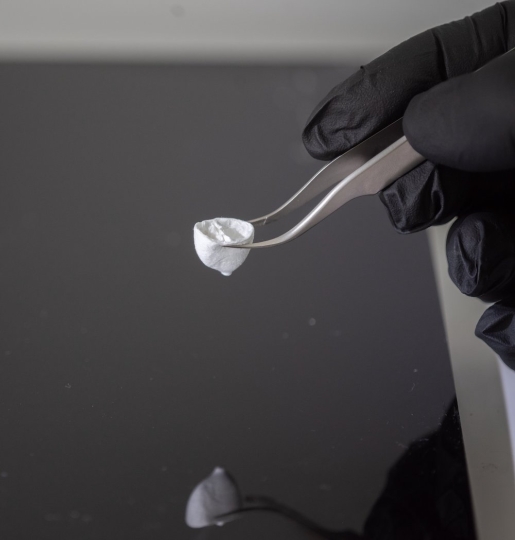
A FRJS spun dual chambered ventricle. (Credit: Disease Biophysics Group/Harvard SEAS)
この進歩は、繊維の積層造形の新しい手法である集束回転ジェット紡糸(FRJS)により実現したもので、直径数マイクロメートルから数百ナノメートルのらせん状に整列した繊維をハイスループットに製造することが可能になりました。
FRJSの最初のステップは、綿菓子製造機のような仕組みだ。液体のポリマー溶液をリザーバーに入れ、装置が回転する際の遠心力によって、小さな開口部から押し出すのである。溶液がタンクから出ると溶媒が蒸発し、ポリマーが固化してファイバーになる。次に、集束した気流によって繊維の配向を制御し、集束装置上に繊維を堆積させる。研究チームは、コレクターの角度を変えたり回転させたりすることで、気流中の繊維が整列し、コレクターのまわりでねじれながら回転し、心臓の筋肉のらせん構造を模倣することを発見した。
繊維の配列は、集電体の角度を変えることで調整することができる。
<関連情報>
- https://www.seas.harvard.edu/news/2022/07/major-step-forward-organ-biofabrication
- https://www.science.org/doi/10.1126/science.abl6395
集束回転ジェットスピンによる心臓のらせん構造-機能関係の再現 Recreating the heart’s helical structure-function relationship with focused rotary jet spinning
Huibin Chang,Qihan Liu,John F. Zimmerman,Keel Yong Lee ,Qianru Jin,Michael M. Peters,Michael Rosnach,Suji Choi,Sean L. Kim ,Herdeline Ann M. Ardoña,Luke A. MacQueen,Christophe O. Chantre,Sarah E. Motta,Elizabeth M. Cordoves,Kevin Kit Parker
Science Published:7 Jul 2022
DOI: 10.1126/science.abl6395
Replicating the structure of a heart
The orientation of matrix fibers in many tissues, including those in the heart, can influence proper function or dysfunction. Chang et al. developed focused rotary jet spinning that enables the deposition of polymeric materials in the form of long, thin fibers with a preferred orientation at the micrometer scale (see the Perspective by Sefton and Simmons). Using this method, the authors were able to fabricate normal hearts that have similar structural properties, but they can also deliberately misalign the fiber orientation to build models of diseased hearts. Once fully constructed, contractile cells are seeded onto the scaffold so that the composite form takes on some of the active properties of a natural heart. This model can also show how the misorientation of specific fibers affects functioning. —MSL
Abstract
Helical alignments within the heart’s musculature have been speculated to be important in achieving physiological pumping efficiencies. Testing this possibility is difficult, however, because it is challenging to reproduce the fine spatial features and complex structures of the heart’s musculature using current techniques. Here we report focused rotary jet spinning (FRJS), an additive manufacturing approach that enables rapid fabrication of micro/nanofiber scaffolds with programmable alignments in three-dimensional geometries. Seeding these scaffolds with cardiomyocytes enabled the biofabrication of tissue-engineered ventricles, with helically aligned models displaying more uniform deformations, greater apical shortening, and increased ejection fractions compared with circumferential alignments. The ability of FRJS to control fiber arrangements in three dimensions offers a streamlined approach to fabricating tissues and organs, with this work demonstrating how helical architectures contribute to cardiac performance.


