2023-06-01 ワシントン大学セントルイス校
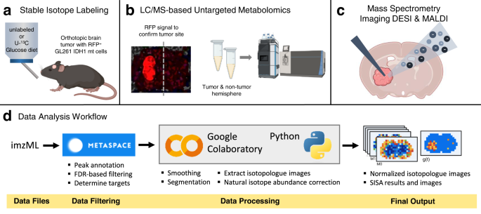
◆この多次元イメージング手法を用いて、彼らは脳がんで特異的に活性化される経路を特定し、治療戦略のヒントを提供しました。この研究は、Nature Communicationsに掲載されました。
◆脳腫瘍モデルを用いて、彼らは「腫瘍生態系」の高解像度地図を作成しました。これにより、脳内のどの部位にどの分子が存在し、それらの分子がどれくらい速く他の物質に変わっているかを把握することができました。
◆この研究は、がん細胞が脳腫瘍内で成長に必要な脂質を作り出すことを明らかにしました。この経路を標的にすることで、病気の進行を遅らせる可能性があります。
<関連情報>
- https://source.wustl.edu/2023/06/cancer-cells-rev-up-synthesis-compared-with-neighbors/
- https://www.nature.com/articles/s41467-023-38403-x
質量分析イメージングを用いて腫瘍の生態系におけるフラックスを定量的にマップする。 Using mass spectrometry imaging to map fluxes quantitatively in the tumor ecosystem
Michaela Schwaiger-Haber,Ethan Stancliffe,Dhanalakshmi S. Anbukumar,Blake Sells,Jia Yi,Kevin Cho,Kayla Adkins-Travis,Milan G. Chheda,Leah P. Shriver & Gary J. Patti
Nature Communications Published:19 May 2023
DOI:https://doi.org/10.1038/s41467-023-38403-x

Abstract
Tumors are comprised of a multitude of cell types spanning different microenvironments. Mass spectrometry imaging (MSI) has the potential to identify metabolic patterns within the tumor ecosystem and surrounding tissues, but conventional workflows have not yet fully integrated the breadth of experimental techniques in metabolomics. Here, we combine MSI, stable isotope labeling, and a spatial variant of Isotopologue Spectral Analysis to map distributions of metabolite abundances, nutrient contributions, and metabolic turnover fluxes across the brains of mice harboring GL261 glioma, a widely used model for glioblastoma. When integrated with MSI, the combination of ion mobility, desorption electrospray ionization, and matrix assisted laser desorption ionization reveals alterations in multiple anabolic pathways. De novo fatty acid synthesis flux is increased by approximately 3-fold in glioma relative to surrounding healthy tissue. Fatty acid elongation flux is elevated even higher at 8-fold relative to surrounding healthy tissue and highlights the importance of elongase activity in glioma.

