2024-05-30 ヒューストン大学(UH)
<関連情報>
- https://uh.edu/news-events/stories/2024/may/05302024-cancer-detection-counting-extracellular-particles.php
- https://www.nature.com/articles/s43856-024-00514-x
癌検出のための単一細胞外小胞のプラズモニック・ナノ・アパーチャー・ラベルフリー・イメージング Plasmonic nano-aperture label-free imaging of single small extracellular vesicles for cancer detection
Nareg Ohannesian,Mohammad Sadman Mallick,Jianzhong He,Yawei Qiao,Nan Li,Simona F. Shaitelman,Chad Tang,Eileen H. Shinn,Wayne L. Hofstetter,Alexei Goltsov,Manal M. Hassan,Kelly K. Hunt,Steven H. Lin & Wei-Chuan Shih
Communications Medicine Published:25 May 2024
DOI:https://doi.org/10.1038/s43856-024-00514-x

Abstract
Background
Small extracellular vesicle (sEV) analysis can potentially improve cancer detection and diagnostics. However, this potential has been constrained by insufficient sensitivity, dynamic range, and the need for complex labeling.
Methods
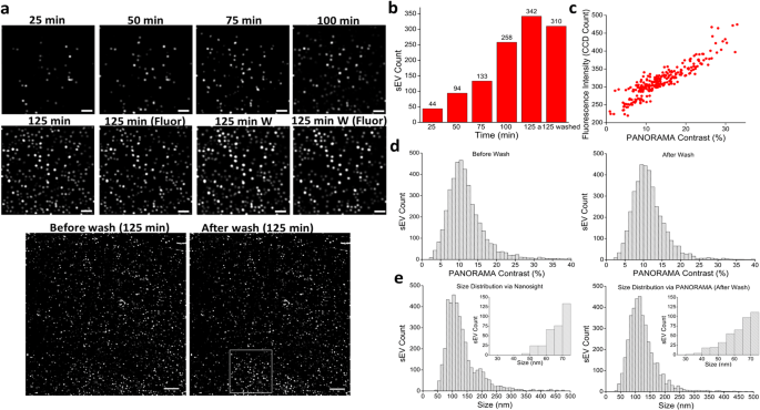
In this study, we demonstrate the combination of PANORAMA and fluorescence imaging for single sEV analysis. The co-acquisition of PANORAMA and fluorescence images enables label-free visualization, enumeration, size determination, and enables detection of cargo microRNAs (miRs).
Results
An increased sEV count is observed in human plasma samples from patients with cancer, regardless of cancer type. The cargo miR-21 provides molecular specificity within the same sEV population at the single unit level, which pinpoints the sEVs subset of cancer origin. Using cancer cells-implanted animals, cancer-specific sEVs from 20 µl of plasma can be detected before tumors were palpable. The level plateaus between 5–15 absolute sEV count (ASC) per µl with tumors ≥8 mm3. In healthy human individuals (N = 106), the levels are on average 1.5 ASC/µl (+/- 0.95) without miR-21 expression. However, for stage I–III cancer patients (N = 205), nearly all (204 out of 205) have levels exceeding 3.5 ASC/µl with an average of 12.2 ASC/µl (±9.6), and a variable proportion of miR-21 labeling among different tumor types with 100% cancer specificity. Using a threshold of 3.5 ASC/µl to test a separate sample set in a blinded fashion yields accurate classification of healthy individuals from cancer patients.
Conclusions
Our techniques and findings can impact the understanding of cancer biology and the development of new cancer detection and diagnostic technologies.
Plain Language Summary
Small extracellular vesicles (sEVs) are tiny particles derived from cells that can be detected in bodily fluids such as blood. Detecting sEVs and analyzing their contents may potentially help us to diagnose disease, for example by observing differences in sEV numbers or contents in the blood of patients with cancer versus healthy people. Here, we combine two imaging methods – our previously developed method PANORAMA and imaging of fluorescence emitted by sEVs—to visualize and count sEVs, determine their size, and analyze their cargo. We observe differences in sEV numbers and cargo in samples taken from healthy people versus people with cancer and are able to differentiate these two populations based on our analysis of sEVs. With further testing, our approach may be a useful tool for cancer diagnosis and provide insights into the biology of cancer and sEVs.

