2025-04-24 北海道大学
北海道大学大学院医学研究院の研究チーム(山崎美和子准教授ら)は、小脳プルキンエ細胞内の小胞体カルシウムストアが機能的に区画化されていることを発見しました。カルシウムセンサータンパク質STIM1が、樹状突起幹の特定の小胞体領域に局在し、IP₃受容体(IP₃R)と一致する一方、リアノジン受容体(RyR)とは重ならないことが明らかになりました。さらに、STIM1をノックダウンした細胞では、IP₃Rを介したカルシウム放出が大きく低下し、STIM1がIP₃感受性カルシウムストアの補充に重要な役割を果たしていることが示されました。この成果は、記憶や学習を支える神経細胞内の情報処理メカニズムの新たな理解につながると期待されます。研究成果は、2025年4月16日付で『The Journal of Neuroscience』に掲載されました。

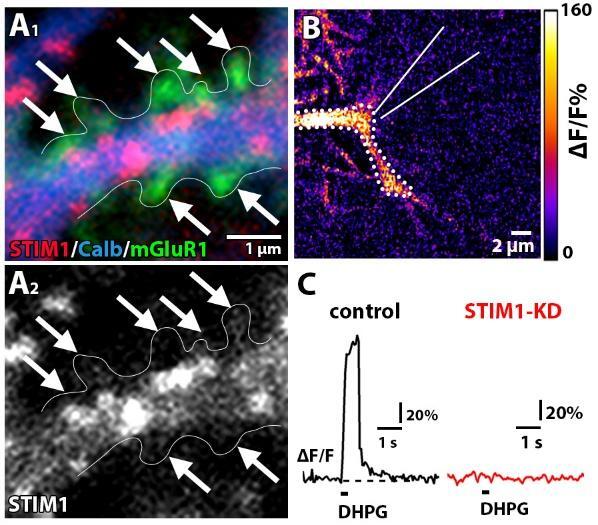
(A)蛍光三重免疫染色により明らかになったマウス小脳プルキンエ細胞の内部のSTIM1の分布。矢印は樹状突起幹から伸びた棘突起を示している。
(B)カルシウムイメージングによるIP3受容体を介したカルシウム上昇の可視化。このカルシウム上昇はSTIM1のノックダウン(STIM1-KD)により消失する(C、右)。
<関連情報>
- https://www.hokudai.ac.jp/news/2025/04/post-1862.html
- https://www.hokudai.ac.jp/news/pdf/250424_pr.pdf
- https://www.jneurosci.org/content/45/16/e1829242025
小胞体カルシウムセンサーSTIM1はマウス小脳プルキンエ細胞の特定の小胞体構造に集積している Preferential Localization of STIM1 to Dendritic Subsurface ER Structures in Mouse Purkinje Cells
Sakyo Nomura, Miwako Yamasaki, Taisuke Miyazaki, Kohtarou Konno and Masahiko Watanabe
The Journal of Neuroscience Published:16 April 2025
DOI:https://doi.org/10.1523/JNEUROSCI.1829-24.2025
Abstract
The endoplasmic reticulum (ER) is the largest intracellular Ca2+ store, serving as the source and sink of intracellular Ca2+. The ER Ca2+ store is continuous yet organized into distinct subcompartments with spatial and functional heterogeneity. In cerebellar Purkinje cells (PCs), glutamatergic inputs trigger Ca2+ release from specific ER domains via inositol 1,4,5-trisphosphate receptors (IP3Rs) or ryanodine receptors (RyRs). Upon ER store depletion, refilling occurs through store-operated Ca2+ entry mediated by stromal interaction molecule-1 (STIM1). Although the significance of STIM1-mediated Ca2+ regulation within PCs is established, STIM1 localization in ER subcompartments in PCs for Ca2+ release and refilling remains elusive. Using validated antibodies, we demonstrated that STIM1 was predominantly localized as intense puncta along dendritic shafts in male and female mice, colocalizing with IP3R1 but not with RyR1. Immunoelectron microscopy revealed that STIM1 was accumulated in the subsurface ER in the dendritic shaft but excluded from those in the dendritic spine, the primary site of metabotropic glutamate receptor 1 (mGluR1)–IP3R-mediated Ca2+ signaling. Ca2+ imaging from control and STIM1-knockdown (STIM1-KD) PCs demonstrated that mGluR1-mediated Ca2+ release is more critically dependent on STIM1 than RyR-mediated Ca2+ release. These findings reveal a spatially organized ER network in PCs, where specialized ER subcompartments differentially regulate Ca2+ release and refilling. These findings suggest that STIM1 preferentially regulates Ca2+ dynamics associated with mGluR1–IP3R signaling, supporting specialized ER subcompartments for Ca2+ release and refilling. These findings highlight the intricate molecular–anatomical organization of dendritic ER Ca2+ signaling in PCs, crucial for synaptic plasticity and motor learning.

