2025-06-09 カリフォルニア大学サンディエゴ校(UCSD)
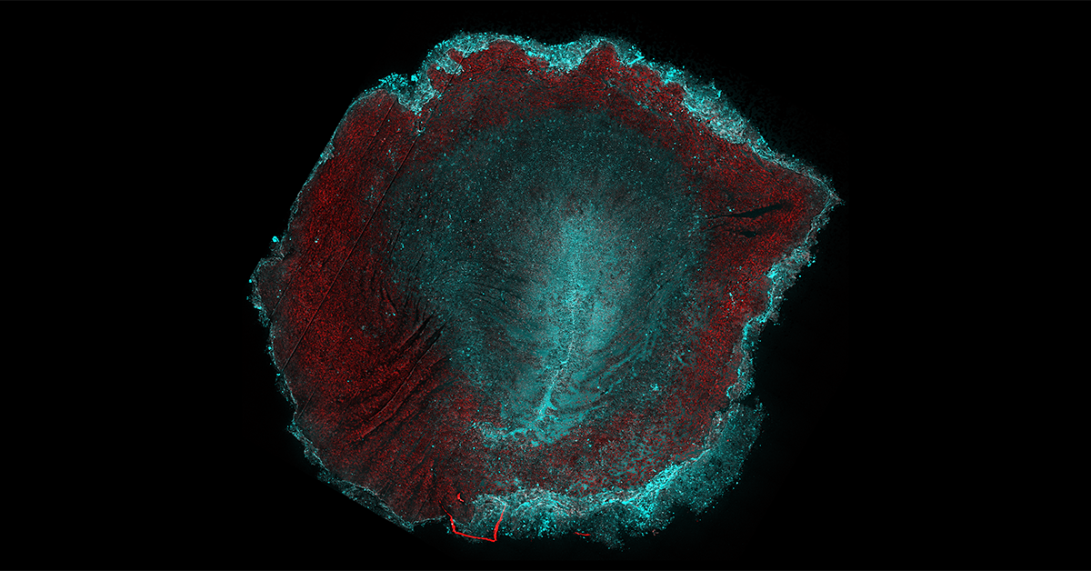
 The experimental image shows two proteins — myosin (cyan) and actin (red) — in a chick embryo during gastrulation, highlighting the main body axes and the dominant force-generating regions (cyan). (cr: Guillermo Serrano Najera)
<関連情報>
- https://today.ucsd.edu/story/understanding-mechanisms-embryonic-cell-behavior
- https://www.nature.com/articles/s41467-025-60249-8
鳥類の胃形成における組織フローと胚形状の制御 Control of tissue flows and embryo geometry in avian gastrulation
Guillermo Serrano Nájera,Alex M. Plum,Ben Steventon,Cornelis J. Weijer & Mattia Serra
Nature Communications Published:04 June 2025
DOI:https://doi.org/10.1038/s41467-025-60249-8
Abstract
Embryonic tissues undergo coordinated flows during avian gastrulation to establish the body plan. Here, we elucidate how the interplay between embryonic and extraembryonic tissues affects the chick embryo’s size and shape. These two distinct geometric changes are each associated with dynamic curves across which trajectories separate (kinematic repellers). Through physical modeling and experimental manipulations of both embryonic and extraembryonic tissues, we selectively eliminate either or both repellers in model and experiments, revealing their mechanistic origins. We find that embryo size is affected by the competition between extraembryonic epiboly and embryonic myosin-driven contraction—which persists when mesoderm induction is blocked. Instead, the characteristic shape change from circular to pear-shaped arises from myosin-driven cell intercalations in the mesendoderm, irrespective of epiboly. These findings elucidate modular mechanisms controlling avian gastrulation flows and provide a mechanistic basis for the independent control of embryo size and shape during development.

