
2025-10-30 国立循環器病研究センター
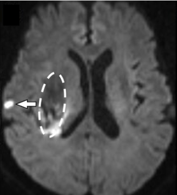
(図1)脳出血(破線囲み部分)を発症した時の“隠れた小さな脳梗塞”(矢印)
<関連情報>
- https://www.ncvc.go.jp/pr/release/pr_49644/
- https://www.neurology.org/doi/10.1212/WNL.0000000000214338
急性脳内出血におけるDWI陽性病変と小血管疾患との相関:コホート研究 DWI-Positive Lesions in Acute Intracerebral Hemorrhage and Their Correlation With Small Vessel Disease A Cohort Study
Satoshi Hosoki, Tomotaka Tanaka, Yuki Kawamura, Sonu M. M. Bhaskar, Soshiro Ogata, Takehito Kuroda, Satoshi Saito, …,and Masafumi Ihara
Neurology Published:October 29, 2025
DOI:https://doi.org/10.1212/WNL.0000000000214338
Abstract
Background and Objectives
Diffusion-weighted imaging (DWI)–positive lesions are identified in 11%–45% of patients with acute intracerebral hemorrhage (ICH); however, their underlying mechanisms and clinical implications remain unclear. Moreover, the prevalence of these lesions before blood pressure lowering remains elusive. The aim of this study was to evaluate the prevalence, time-dependent changes, and associations of DWI-positive lesions with small vessel disease (SVD) markers and clinical outcomes in patients with acute ICH.
Methods
This retrospective cohort study analyzed data from the National Cerebral and Cardiovascular Center stroke registry. Overall, 872 patients with acute ICH who underwent MRI between January 2015 and January 2021 were evaluated. The patients were characterized based on whether MRI was performed before or after acute-phase blood pressure lowering. Multivariable logistic regression was used to assess the correlation between DWI-positive lesions and factors related to blood pressure reduction and time from ICH onset to imaging, as well as correlation with SVD markers. Furthermore, the relationship between DWI-positive lesions and unfavorable clinical outcomes (modified Rankin Scale scores 3–6 at 90 days) was examined.
Results
Among 872 patients, 114 patients (13.1%) exhibited DWI-positive lesions (mean age: 71.5 ± 12.9 years; female, 36.0%). The number of patients with DWI-positive lesions was lower among those who underwent MRI before acute-phase blood pressure lowering (45/444 patients [10.1%]) than among those who underwent MRI after blood pressure lowering (69/428 patients [16.1%]). Multivariable analysis revealed that DWI-positive lesions were significantly associated with time from ICH onset to imaging (adjusted odds ratio [aOR], 1.41; 95% CI 1.08–1.84), but not with blood pressure lowering (aOR, 0.92; 95% CI 0.51–1.65). DWI-positive lesions were also strongly associated with SVD markers and higher SVD severity scores. Furthermore, DWI-positive lesions were linked to unfavorable outcomes at 90 days (aOR, 1.70; 95% CI 1.04–2.80).
Discussion
DWI-positive lesions are observed before blood pressure lowering, and their frequency increases over time. Their association with SVD markers highlights the role of advanced SVD in lesion formation. These lesions may worsen clinical outcomes, can potentially be helpful as prognostic imaging biomarkers, and may help guide optimal acute ICH management.

")