
")
2026-03-20 マックス・プランク研究所
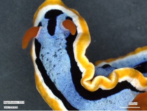
Nudibranchs such as Chromodoris annae stand out for their vibrant colours and varied shapes. C. annae is found in the Indo-Pacific region around Malaysia, Indonesia, the Philippines and the Marshall Islands.© Samuel Humphrey/MPI of Colloids and Interfaces
<関連情報>
- https://www.mpg.de/26287823/impressionist-sea-slugs
- https://www.pnas.org/doi/10.1073/pnas.2525419123
ウミウシの体色の多様性は、グアニン光子構造「ピクセル」という共通の物理的基盤に基づいている Nudibranch color diversity shares a common physical basis in guanine photonic structure ‘pixels’
Samuel Humphrey, Xianglian He, Tobias Priemel, +5 , and Silvia Vignolini
Proceedings of the National Academy of Sciences Published:March 17, 2026
DOI:https://doi.org/10.1073/pnas.2525419123
Significance
Nudibranchs are an extraordinarily diverse group of marine animals, renowned for their dazzling range of colors and striking patterns. While their pigmentary coloration is well understood, so far, structural coloration has been largely overlooked. In this work, we present a comparative analysis of structural coloration across nudibranch species from benthic and coral reef environments, demonstrating that guanine-based nanostructures are a widespread motif responsible for many of the brilliant colors within the dorid and aeolid groups. These nanostructures produce a unique optical mechanism, whereby matte coloration across the entire visible range can be achieved using hierarchical multilayer architectures. Microscopically, the color appears pixelated, and individual multilayers reflect a specific wavelength; these reflections are mixed at the macroscopic level, generating vibrant hues.
Abstract
Nudibranchs are well known for their bright and diverse color patterns. This coloration is typically a form of aposematism, warning predators against toxic compounds sequestered from their prey and weaponized as a form of defense. Although many of the hues in nudibranchs were thought to be of pigmentary origin, here we show, using a combination of white light and Raman microspectroscopy, that hierarchically organized micron-scale guanine multilayer structures are responsible for many of these colors. Such architectures are widespread across the dorid and aeolid groups and are responsible for a striking array of angular-independent structural colors. By using cryogenic focused ion beam (cryo-FIB) SEM tomography, we were able to access the complex 3D organization of the guanine nanoplatelets responsible for the strong blue coloration of Chromodoris annae. We propose that the multilayer organization of guanine platelets with varying orientations across the tissue and their micron-scale size offers a particularly effective strategy for producing diverse optical effects. The macroscopic angular independent color results from individual multilayers which we describe as “pixels”, these “pixels” reflect light at a wavelength governed by their interlayer spacing and guanine platelet thickness. The macroscopic hue can be spectrally tuned by altering the statistical distribution of pixels with each color, while the angular dependence of color can be changed through the relative orientation of the multilayer stacks and their size, allowing for a single structural motif to generate a broad palette of optical appearances.
")
")