2026-09-29 東京科学大学
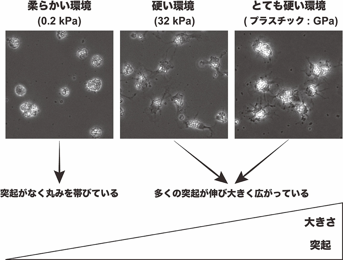
図1. 肝星細胞の活性化初期における硬さ別の形態変化。柔らかい環境では、肝星細胞の活性化初期の形態変化が抑えられている。
<関連情報>
- https://www.isct.ac.jp/ja/news/gm2qk7n873j0
- https://link.springer.com/article/10.1007/s12072-026-11141-0
細胞外マトリックスの硬さが肝星細胞活性化における初期の転写経路を形成する Matrix stiffness shapes early transcriptional trajectories during hepatic stellate cell activation
Kento Inada, Masato Miyoshi, Sei Kakinuma, Keiya Watakabe, Tomohiro Mochida, Taro Shimizu, Jun Tsuchiya, Tsubasa Nobusawa, Shun Kaneko, Fukiko Kawai-Kitahata, Miyako Murakawa, Sayuri Nitta, Mina Nakagawa, Yasuhiro Asahina & Ryuichi Okamoto
Hepatology International Published:01 September 2026
DOI:https://doi.org/10.1007/s12072-026-11141-0
Abstract
Background
Matrix stiffness increases during fibrosis and drives hepatic stellate cell (HSC) activation through YAP-dependent mechanotransduction. Although recent studies have revealed HSC heterogeneity beyond classical quiescent and fully activated states, how physiological-range stiffness influences activation trajectories underlying this heterogeneity remains unclear. This study aimed to determine how substrate stiffness shapes the transcriptomic profiles and biases activation trajectories.
Methods
We performed time-resolved single-cell RNA sequencing of primary mouse HSCs cultured on stiffness-tunable polydimethylsiloxane (PDMS) substrates (Soft, 0.2 kPa; Hard, 32 kPa), using tissue culture polystyrene (TCPS) as a conventional baseline. In vitro findings were anchored to public in vivo datasets from mouse and human liver injury.
Results
Matrix stiffness altered HSC morphology and transcriptional state, with softer substrates promoting a rounder phenotype and reducing the expression of activation markers, particularly during early activation. Pseudotime and RNA velocity analyses also revealed different early trajectories associated with matrix stiffness. Under the soft condition, HSCs exhibited an expanded quiescent-to-initiatory transitional state characterized by Nrf2-related features and regeneration-associated factors, including Hgf and Rspo3. In contrast, the hard condition induced a gene signature, which was upregulated from early stages of HSC activation onward, localized to fibrotic septa in human liver and correlated with cirrhosis severity in an HCV patient cohort.
Conclusion
Matrix stiffness shapes early HSC activation trajectories toward transcriptional programs associated with regeneration or fibrosis. These findings support stiffness-controlled culture systems as improved models of HSC activation and provide a framework for regenerative and antifibrotic strategies.