一次視覚野の大きさと、視覚情報の処理に特化した脳組織の量から、私たちがどれだけよく見えるかを予測できることが、新しい研究で明らかに。 The size of our primary visual cortex and the amount of brain tissue we have dedicated to processing visual information can predict how well we can see, a new study shows.
2022-06-13 ニューヨーク大学 (NYU)
研究チームは、機能的磁気共鳴画像法(fMRI)を用いて、20人以上のヒトの一次視覚野(V1)の大きさを測定した。さらに、視野内の異なる位置(左、右、上、下)からの視覚情報を処理するために、V1組織の量も測定された。
また、V1測定と同じ視野の位置で、視覚の質を評価するための課題も実施した。この課題では、コンピュータ画面上に表示されたパターンの向きを識別し、「コントラスト感度」、つまり画像間の区別をする能力を測定した。
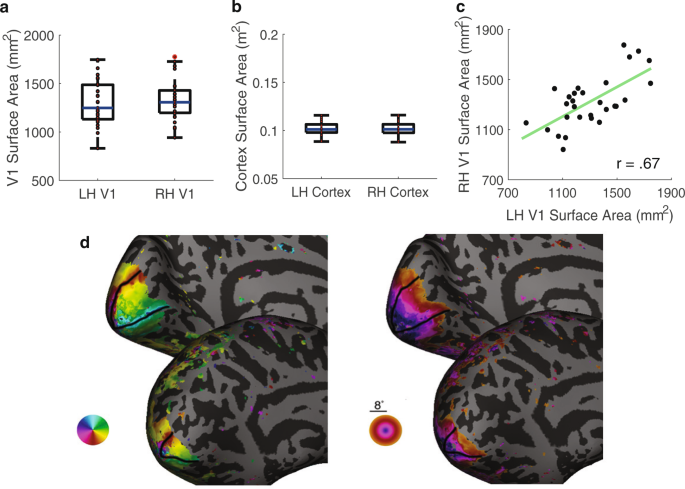
その結果、V1の表面積の違いが、コントラスト感度の測定値を予測することがわかった。まず、V1の面積が大きい人は、小さい人よりも全体的にコントラスト感度が高かった(表面積が最大の人は1,776平方ミリメートル、最小の人は832平方ミリメートルであった)。第二に、視野内の特定の領域からの視覚情報を処理する皮質組織がV1に多い人は、同じ領域に専念する皮質組織が少ない人に比べて、その領域でのコントラスト感度が高いということである。第三に、参加者全体で、固定点から等距離にある別の場所(例:上)よりも特定の場所(例:左)でコントラスト感度が高いことは、それぞれ皮質組織の多い領域または少ない領域に対応する。
<関連情報>
- https://www.nyu.edu/about/news-publications/news/2022/june/neuroscientists-find-new-factors-behind-better-vision-.html
- https://www.nature.com/articles/s41467-022-31041-9
ヒト一次視覚野の個人差と視野周辺のコントラスト感度の関連性 Linking individual differences in human primary visual cortex to contrast sensitivity around the visual field
Marc M. Himmelberg,Jonathan Winawer &Marisa Carrasco
Nature Communications Published:13 June 2022
DOI:https://doi.org/10.1038/s41467-022-31041-9

Abstract
A central question in neuroscience is how the organization of cortical maps relates to perception, for which human primary visual cortex (V1) is an ideal model system. V1 nonuniformly samples the retinal image, with greater cortical magnification (surface area per degree of visual field) at the fovea than periphery and at the horizontal than vertical meridian. Moreover, the size and cortical magnification of V1 varies greatly across individuals. Here, we used fMRI and psychophysics in the same observers to quantify individual differences in V1 cortical magnification and contrast sensitivity at the four polar angle meridians. Across observers, the overall size of V1 and localized cortical magnification positively correlated with contrast sensitivity. Moreover, greater cortical magnification and higher contrast sensitivity at the horizontal than the vertical meridian were strongly correlated. These data reveal a link between cortical anatomy and visual perception at the level of individual observer and stimulus location.

