3億1900万年前の魚の化石から、動物がどのように進化して現在のような生物になったのか、新たな知見が得られた A 319-million-year-old fossilised fish reveals fresh insight into how animals evolved into the creatures we see today
2023-01-31 バーミンガム大学
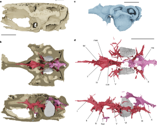
◆X線を使って内部の特徴を明らかにするCTスキャンにより、この生物の頭蓋骨には、およそ1インチの長さの脳と脳神経があることがわかりました。
◆バーミンガム大学(英国)とミシガン大学(米国)の研究者らは、この発見が現在生きている魚類の主要なグループである光線性魚類の神経解剖学と初期の進化を知る窓を開くと考えています。
◆この研究成果は、背骨のある動物の化石に柔らかい部分が保存されていることに新たな光を当てたもので、『Nature』誌に発表された。博物館に収蔵されている動物の化石のほとんどは、骨や歯、貝殻などの体の硬い部分から形成されている。
◆バーミンガム大学の主任研究員Sam Giles氏は、次のようにコメントしています。「今回、脊椎動物の脳が立体的に保存されているという予想外の発見により、エイヒレ科魚類の神経解剖学について驚くべき洞察を得ることができました。この発見は、現生種の脳が示唆するよりも複雑な脳の進化パターンを教えてくれ、現在の硬骨魚類がいつ、どのように進化してきたかをより明確にすることができます。
◆”現存する魚類との比較から、コッコケファルスの脳は、チョウザメやパドルフィッシュの脳に最も似ていることが分かりました。”チョウザメやパドルフィッシュは、3億年以上前に他の全ての現存する鱏魚類から分岐したため「始祖魚」と呼ばれることが多い魚類です。
◆分析されたCTスキャンされた脳は、Coccocephalus wildiのもので、河口で泳ぎ、小さな甲殻類、水生昆虫、頭足類(今日イカ、タコ、イカを含むグループ)を食べていたと思われる、鯛とほぼ同じサイズの初期の光線性魚のものです。光線魚類は、背骨と鰭を光線と呼ばれる骨の棒で支えている。
◆通常、脳などの軟組織はすぐに腐敗してしまい、化石になることはほとんどありません。しかし、この魚が死んだとき、脳と脳神経の軟組織は、化石化の過程で高密度の鉱物と交換され、その三次元構造が精巧に保存された。
◆この化石は、エイ科魚類の脳の特徴的な機能が進化する前の時代を捉えており、この特徴がいつ進化したかを示す指標となる。
<関連情報>
- https://www.birmingham.ac.uk/news/2023/319-million-year-old-fossilised-fish-illuminates-backboned-animals-brain-evolution
- https://www.nature.com/articles/s41586-022-05666-1
エイヒレ科魚類の脳の例外的な化石保存とその進化 Exceptional fossil preservation and evolution of the ray-finned fish brain
Rodrigo T. Figueroa,Danielle Goodvin,Matthew A. Kolmann,Michael I. Coates,Abigail M. Caron,Matt Friedman & Sam Giles
Nature Published:01 February 2023
DOI:https://doi.org/10.1038/s41586-022-05666-1

Abstract
Brain anatomy provides key evidence for the relationships between ray-finned fishes1, but two major limitations obscure our understanding of neuroanatomical evolution in this major vertebrate group. First, the deepest branching living lineages are separated from the group’s common ancestor by hundreds of millions of years, with indications that aspects of their brain morphology—like other aspects of their anatomy2,3—are specialized relative to primitive conditions. Second, there are no direct constraints on brain morphology in the earliest ray-finned fishes beyond the coarse picture provided by cranial endocasts: natural or virtual infillings of void spaces within the skull4,5,6,7,8. Here we report brain and cranial nerve soft-tissue preservation in Coccocephalus wildi, an approximately 319-million-year-old ray-finned fish. This example of a well-preserved vertebrate brain provides a window into neural anatomy deep within ray-finned fish phylogeny. Coccocephalus indicates a more complicated pattern of brain evolution than suggested by living species alone, highlighting cladistian apomorphies1 and providing temporal constraints on the origin of traits uniting all extant ray-finned fishes1,9. Our findings, along with a growing set of studies in other animal groups10,11,12, point to the importance of ancient soft tissue preservation in understanding the deep evolutionary assembly of major anatomical systems outside of the narrow subset of skeletal tissues13,14,15.


