2023-10-24 ワシントン大学セントルイス校
◆研究者は、C-Cケモカイン受容体2の発現を持つ炎症性免疫細胞を標識するためのラジオトレーサーを開発し、心臓発作を経験した患者で陽電子放射線断層撮影を使用してこれらの細胞を視覚化しました。この研究は、免疫調節療法を受けるべき個人を特定する上で役立つ可能性があります。
<関連情報>
- https://source.wustl.edu/2023/10/noninvasive-technique-helps-visualize-inflammatory-cells-in-human-heart/
- https://www.nature.com/articles/s44161-023-00335-6
ヒトST上昇型心筋梗塞におけるCCR2イメージング CCR2 imaging in human ST-segment elevation myocardial infarction
Kory J. Lavine,Deborah Sultan,Hannah Luehmann,Lisa Detering,Xiaohui Zhang,Gyu Seong Heo,Xiuli Zhang,Michelle Hoelscher,Kitty Harrison,Christophe Combadière,Richard Laforest,Daniel Kreisel,Pamela K. Woodard,Steven L. Brody,Robert J. Gropler & Yongjian Liu
Nature Cardiovascular Research Published:21 September 2023
DOI:https://doi.org/10.1038/s44161-023-00335-6

Abstract
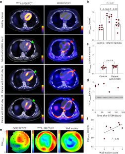
Among the diverse populations of myeloid cells that reside within the healthy and diseased heart, C-C chemokine receptor type 2 (CCR2) is specifically expressed on inflammatory populations of monocytes and macrophages that contribute to the development and progression of heart failure1,2,3,4. Here, we evaluated a peptide-based imaging probe (64Cu-DOTA-ECL1i) that specifically recognizes CCR2+ monocytes and macrophages for human cardiac imaging. Compared to healthy controls, 64Cu-DOTA-ECL1i heart uptake was increased in individuals after acute myocardial infarction, predominately localized within the infarct area, and was associated with impaired myocardial wall motion. These findings establish the feasibility of molecular imaging of CCR2 expression to visualize inflammatory monocytes and macrophages in the injured human heart.


