従来の方法よりも効果的かつ効率的なこの新しいプロセスは、がん診断だけでなく、他の研究分野にも大きな影響を与える可能性があります。 The new process, which is more effective and efficient than conventional methods, has the potential to significantly impact cancer diagnostics as well as other fields of research.
2022-07-14 ブラウン大学
新しいプロセスでは、2枚の平行平板電極の間にある液体で満たされた容器に組織生検が入れられる。酵素の代わりに電界の変動が加えられて、液体内に反対方向の力が発生する。この力によって、組織細胞は一方向に動き、次に反対方向に動き、互いにきれいに分離する(解離する)。
この新しいプロセスは、わずか5分で生検組織の解離をもたらしました。これは、TripathiとWelchが以前の研究で説明した主要な酵素的、機械的技術よりも3倍速いスピードです。
このアプローチでは、細胞の生存率、形態、細胞周期の進行を維持したまま、組織を単細胞に良好に解離することができ、単細胞の直接分析のための組織試料の前処理に有用であることが示唆された。
<関連情報>
- https://www.brown.edu/news/2022-07-14/single-cell-isolation
- https://www.nature.com/articles/s41598-022-13068-6
電場による組織の迅速かつ効率的な解離 生きた単一細胞への変換 Electric-field facilitated rapid and efficient dissociation of tissues Into viable single cells
E. Celeste Welch,Harry Yu,Gilda Barabino,Nikos Tapinos & Anubhav Tripathi
Scientific Reports Published:24 June 2022
DOI:https://doi.org/10.1038/s41598-022-13068-6

Abstract
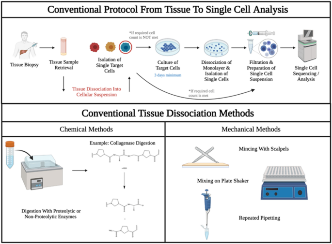
Single-Cell Analysis is a growing field that endeavors to obtain genetic profiles of individual cells. Disruption of cell–cell junctions and digestion of extracellular matrix in tissues requires tissue-specific mechanical and chemical dissociation protocols. Here, a new approach for dissociating tissues into constituent cells is described. Placing a tissue biopsy core within a liquid-filled cavity and applying an electric field between two parallel plate electrodes facilitates rapid dissociation of complex tissues into single cells. Different solution compositions, electric field strengths, and oscillation frequencies are investigated experimentally and with COMSOL Multiphysics. The method is compared with standard chemical and mechanical approaches for tissue dissociation. Treatment of tissue samples at 100 V/cm 1 kHz facilitated dissociation of 95 ± 4% of biopsy tissue sections in as little as 5 min, threefold faster than conventional chemical–mechanical techniques. The approach affords good dissociation of tissues into single cells while preserving cell viability, morphology, and cell cycle progression, suggesting utility for sample preparation of tissue specimens for direct Single-Cell Analysis.

