私たちの「見え方」を解明する新しい研究 New Study Enhances Understanding of How We See
2022-10-04 ニューヨーク大学 (NYU)

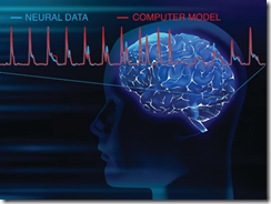
Rapid fluctuations in neural activity in the human visual cortex, analyzed by a team of neuroscientists, are well predicted by a computer model, as shown above. Brain image by rawpixel.com.
特定の現象(発作に伴う脳活動)を測定するために、電極を埋め込んだボランティアのてんかん患者を対象に調査を行った。
患者は、病院のベッドサイドに置かれたノートパソコンで映像を見ながら研究に参加し、神経科学者たちが珍しい新しい測定を行えるようにした。
脳活動の測定結果から、神経反応を説明するために開発された既存の計算モデルが人間の脳にも適用可能であることが示された。これらのモデルは、人間以外の霊長類を対象とした過去の研究に基づいて、人間以外の神経活動をマッピングしたものである。
<関連情報>
- https://www.nyu.edu/about/news-publications/news/2022/october/rare-electrical-recordings-of-the-human-brain-give-detailed-pict.html
- https://www.jneurosci.org/content/early/2022/09/28/JNEUROSCI.0690-21.2022.abstract
大規模なヒトの観察者におけるV1、V2、V3マップの表面積のばらつき Variability of the Surface Area of the V1, V2, and V3 Maps in a Large Sample of Human Observers
Noah C. Benson, Jennifer M. D. Yoon, Dylan Forenzo, Stephen A. Engel, Kendrick N. Kay and Jonathan Winawer
Journal of Neuroscience Published:30 September 2022
DOI: https://doi.org/10.1523/JNEUROSCI.0690-21.2022
Abstract
How variable is the functionally-defined structure of early visual areas in human cortex and how much variability is shared between twins? Here we quantify individual differences in the best understood functionally-defined regions of cortex: V1, V2, V3. The Human Connectome Project 7T Retinotopy Dataset includes retinotopic measurements from 181 subjects (109 female, 72 male), including many twins. We trained four “anatomists” to manually define V1-V3 using retinotopic features. These definitions were more accurate than automated anatomical templates and showed that surface areas for these maps varied more than three-fold across individuals. This three-fold variation was little changed when normalizing visual area size by the surface area of the entire cerebral cortex. In addition to varying in size, we find that visual areas vary in how they sample the visual field. Specifically, the cortical magnification function differed substantially among individuals, with the relative amount of cortex devoted to central vision varying by more than a factor of 2. To complement the variability analysis, we examined the similarity of visual area size and structure across twins. Whereas the twin sample sizes are too small to make precise heritability estimates (50 monozygotic pairs, 34 dizygotic pairs), they nonetheless reveal high correlations, consistent with strong effects of the combination of shared genes and environment on visual area size. Collectively, these results provide the most comprehensive account of individual variability in visual area structure to date, and provide a robust population benchmark against which new individuals and developmental and clinical populations can be compared.
SIGNIFICANCE STATEMENT:
Areas V1, V2, and V3 are among the best studied functionally-defined regions in human cortex. Using the largest retinotopy dataset to date, we characterized the variability of these regions across individuals and the similarity between twin pairs. We find that the size of visual areas varies dramatically (up to 3.5x) across healthy young adults, far more than the variability of the cerebral cortex size as a whole. Much of this variability appears to arise from inherited factors, as we find very high correlations in visual area size between monozygotic twin-pairs, and lower but still substantial correlations between dizygotic twin pairs. These results provide the most comprehensive assessment of how functionally defined visual cortex varies across the population to date.

