カリフォルニア大学サンディエゴ校のエンジニアが、運動中でも使える心臓画像用の強力な新超音波センサーシステムの開発を主導 UC San Diego engineers lead development of a powerful new ultrasound sensor system for cardiac imaging that even works during a workout
2023-01-25 カリフォルニア大学サンディエゴ校(UCSD)
◆このプロジェクトのリーダーであるカリフォルニア大学サンディエゴ校のSheng Xu教授(ナノエンジニアリング)は、「目標は、より多くの人々が超音波検査を利用できるようにすることです」と語っています。現在、心臓の超音波検査である心エコー図は、高度な訓練を受けた技術者とかさばる装置を必要とします。「この技術によって、誰でも外出先で超音波画像を利用できるようになります」とXuは述べています。カスタムAIアルゴリズムのおかげで、この装置は心臓がどれだけ血液を送り出しているかを測定することができる。この研究は、1月25日発行の『Nature』誌に掲載されました。
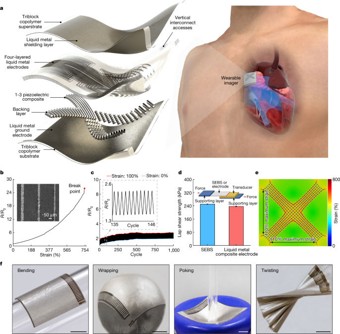
◆新システムは、人間の皮膚と同じ柔らかさのウェアラブルパッチで情報を収集し、最適な装着ができるよう設計されています。パッチは1.9cm(長さ)×2.2cm(幅)×0.09cm(厚さ)と、切手ほどの大きさです。このパッチは、超音波を送受信し、心臓の構造をリアルタイムで連続的に画像化するために使用されます。この超音波パッチは柔らかく伸縮自在で、運動中でも人の皮膚によく密着する。
◆このシステムは、超音波を使って心臓の左心室を別々のバイ・プレーン・ビューで検査することができ、従来よりも臨床的に有用な画像を生成することができます。ユースケースとして、臨床現場で使用されている硬くて扱いにくい機器では不可能な、運動中の心臓の撮影を実演しました。
◆心臓の性能は、1回の拍動で心臓が送り出す血液量である一回拍出量、心臓の左心室から送り出される血液の割合である駆出率、1分間に送り出される血液量である心拍出量の3要素で特徴付けられる。Xu氏のチームは、AIによる連続的な自動処理を促進するアルゴリズムを開発しました。
<関連情報>
- https://today.ucsd.edu/story/cardiac-imaging-on-the-go
- https://www.nature.com/articles/s41586-022-05498-z
ウェアラブル心臓超音波イメージャ A wearable cardiac ultrasound imager
Hongjie Hu,Hao Huang,Mohan Li,Xiaoxiang Gao,Lu Yin,Ruixiang Qi,Ray S. Wu,Xiangjun Chen,Yuxiang Ma,Keren Shi,Chenghai Li,Timothy M. Maus,Brady Huang,Chengchangfeng Lu,Muyang Lin,Sai Zhou,Zhiyuan Lou,Yue Gu,Yimu Chen,Yusheng Lei,Xinyu Wang,Ruotao Wang,Wentong Yue,Xinyi Yang,Yizhou Bian,Jing Mu,Geonho Park,Shu Xiang,Shengqiang Cai,Paul W. Corey,Joseph Wang & Sheng Xu
Nature Published:25 January 2023
DO:Ihttps://doi.org/10.1038/s41586-022-05498-z

Abstract
Continuous imaging of cardiac functions is highly desirable for the assessment of long-term cardiovascular health, detection of acute cardiac dysfunction and clinical management of critically ill or surgical patients1,2,3,4. However, conventional non-invasive approaches to image the cardiac function cannot provide continuous measurements owing to device bulkiness5,6,7,8,9,10,11, and existing wearable cardiac devices can only capture signals on the skin12,13,14,15,16. Here we report a wearable ultrasonic device for continuous, real-time and direct cardiac function assessment. We introduce innovations in device design and material fabrication that improve the mechanical coupling between the device and human skin, allowing the left ventricle to be examined from different views during motion. We also develop a deep learning model that automatically extracts the left ventricular volume from the continuous image recording, yielding waveforms of key cardiac performance indices such as stroke volume, cardiac output and ejection fraction. This technology enables dynamic wearable monitoring of cardiac performance with substantially improved accuracy in various environments.


