2023-07-27 ワシントン大学セントルイス校
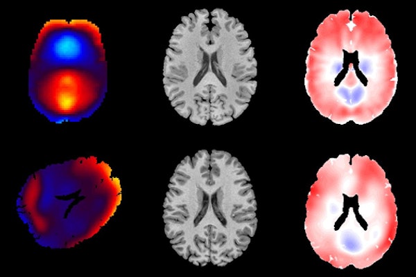
◆インパルス応答とハーモニック応答を測定し、MRE(MRエラストグラフィ)という非侵襲的な技術を使用して脳の動きを観察しました。MREは脳の衝撃に対する応答を示すのに有用で、保護装置の設計や外傷性脳損傷の研究に役立つとされています。
◆彼らの研究により、脳の変形パターンがMREで生成されるものと非常に類似していることがわかりました。この結果は、脳の生体力学モデルの評価と改善に貢献し、外傷性脳損傷の社会的負担を軽減する可能性があります。
<関連情報>
- https://source.wustl.edu/2023/07/brain-movement-measured-for-clues-to-prevent-reduce-injury/
- https://asmedigitalcollection.asme.org/biomechanical/article/145/8/081006/1164116/Comparison-of-Deformation-Patterns-Excited-in-the
頭蓋骨の調和運動と衝動運動により生体内でヒト脳に励起される変形パターンの比較 Comparison of Deformation Patterns Excited in the Human Brain In Vivo by Harmonic and Impulsive Skull Motion
Jordan D. Escarcega,,Andrew K. Knutsen,,Ahmed A. Alshareef,,Curtis L. Johnson,,Ruth J. Okamoto,,Dzung L. Pham,,Philip V. Bayly
Journal of Biomechanical Engineering Published:June 30, 2023
DOI:https://doi.org/10.1115/1.4062809
Abstract
Noninvasive measurements of brain deformation in human participants in vivo are needed to develop models of brain biomechanics and understand traumatic brain injury (TBI). Tagged magnetic resonance imaging (tagged MRI) and magnetic resonance elastography (MRE) are two techniques to study human brain deformation; these techniques differ in the type of motion and difficulty of implementation. In this study, oscillatory strain fields in the human brain caused by impulsive head acceleration and measured by tagged MRI were compared quantitatively to strain fields measured by MRE during harmonic head motion at 10 and 50 Hz. Strain fields were compared by registering to a common anatomical template, then computing correlations between the registered strain fields. Correlations were computed between tagged MRI strain fields in six participants and MRE strain fields at 10 Hz and 50 Hz in six different participants. Correlations among strain fields within the same experiment type were compared statistically to correlations from different experiment types. Strain fields from harmonic head motion at 10 Hz imaged by MRE were qualitatively and quantitatively similar to modes excited by impulsive head motion, imaged by tagged MRI. Notably, correlations between strain fields from 10 Hz MRE and tagged MRI did not differ significantly from correlations between strain fields from tagged MRI. These results suggest that low-frequency modes of oscillation dominate the response of the brain during impact. Thus, low-frequency MRE, which is simpler and more widely available than tagged MRI, can be used to illuminate the brain’s response to head impact.


