2023-12-06 ワシントン大学セントルイス校
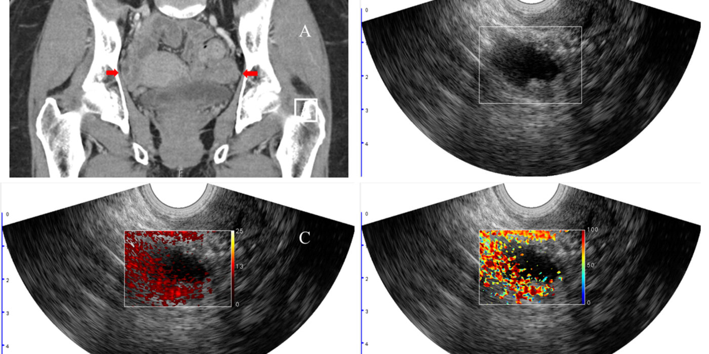
 Quing Zhu, working with Matthew Powell, MD, and Cary Siegel, MD, from the School of Medicine, developed a new imaging method to better diagnose lesions in the ovaries and adjacent adnexa that may help to avoid unnecessary surgeries. This image shows a woman with pelvic mass and BRCA1 mutation. The arrows point to the right and left adnexa. Co-registered ultrasound shows a cyst with solid component in the right adnexa (B). Image C shows the relative total hemoglobin in the lesion, and image D shows the oxygen saturation, both of which are important predictors of malignancy. Pathology revealed a stage I high-grade serous carcinoma of the right ovary and fallopian tube. (Image: Zhu lab)
◆フォトアコースティックイメージングは、近赤外線光を使用して組織を照らし、良性と悪性の病変を区別するための補完的な診断画像データを提供します。研究では、悪性病変は通常、腫瘍血管新生によるヘモグロビン濃度の上昇と、高い腫瘍代謝による低い酸素飽和度を示すことが分かりました。
◆研究者は68人の患者で行われた臨床研究で、この技術を適用し、患者の相対的な総ヘモグロビンや酸素飽和度などの要因を組み合わせたモデルが、診断モデルの性能を測定する指標であるAUCが0.97であったことを示しました。
<関連情報>
- https://source.wustl.edu/2023/12/photoacoustic-imaging-improves-diagnostic-accuracy-of-cancerous-ovarian-lesions/
- https://obgyn.onlinelibrary.wiley.com/doi/10.1002/uog.27452
超音波検査によるO-RADSリスク評価を改善するための光超音波イメージングを用いた付属器病変の特性評価 Characterization of adnexal lesions using photoacoustic imaging to improve sonographic O-RADS risk assessment
Q. Zhu, H. Luo, W. D. Middleton, M. Itani, I. S. Hagemann, A. R. Hagemann, M. J. Hoegger, P. H. Thaker, L. M. Kuroki, C. K. McCourt, D. G. Mutch, M. A. Powell, C. L. Siegel
Ultrasound in Obstetrics and Gynecology Published: 22 August 2023
DOI:https://doi.org/10.1002/uog.27452
Abstract
Objective
To assess the impact of photoacoustic imaging (PAI) on the assessment of ovarian/adnexal lesion(s) of different risk categories using the sonographic ovarian-adnexal imaging-reporting-data system (O-RADS) in women undergoing planned oophorectomy.
Method
This prospective study enrolled women with ovarian/adnexal lesion(s) suggestive of malignancy referred for oophorectomy. Participants underwent clinical ultrasound (US) examination followed by coregistered US and PAI prior to oophorectomy. Each ovarian/adnexal lesion was graded by two radiologists using the US O-RADS scale. PAI was used to compute relative total hemoglobin concentration (rHbT) and blood oxygenation saturation (%sO2) colormaps in the region of interest. Lesions were categorized by histopathology into malignant ovarian/adnexal lesion, malignant Fallopian tube only and several benign categories, in order to assess the impact of incorporating PAI in the assessment of risk of malignancy with O-RADS. Malignant and benign histologic groups were compared with respect to rHbT and %sO2 and logistic regression models were developed based on tumor marker CA125 alone, US-based O-RADS alone, PAI-based rHbT with %sO2, and the combination of CA125, O-RADS, rHbT and %sO2. Areas under the receiver-operating-characteristics curve (AUC) were used to compare the diagnostic performance of the models.
Results
There were 93 lesions identified on imaging among 68 women (mean age, 52 (range, 21–79) years). Surgical pathology revealed 14 patients with malignant ovarian/adnexal lesion, two with malignant Fallopian tube only and 52 with benign findings. rHbT was significantly higher in malignant compared with benign lesions. %sO2 was lower in malignant lesions, but the difference was not statistically significant for all benign categories. Feature analysis revealed that rHbT, CA125, O-RADS and %sO2 were the most important predictors of malignancy. Logistic regression models revealed an AUC of 0.789 (95% CI, 0.626–0.953) for CA125 alone, AUC of 0.857 (95% CI, 0.733–0.981) for O-RADS only, AUC of 0.883 (95% CI, 0.760–1) for CA125 and O-RADS and an AUC of 0.900 (95% CI, 0.815–0.985) for rHbT and %sO2 in the prediction of malignancy. A model utilizing all four predictors (CA125, O-RADS, rHbT and %sO2) achieved superior performance, with an AUC of 0.970 (95% CI, 0.932–1), sensitivity of 100% and specificity of 82%.
Conclusions
Incorporating the additional information provided by PAI-derived rHbT and %sO2 improves significantly the performance of US-based O-RADS in the diagnosis of adnexal lesions. © 2023 International Society of Ultrasound in Obstetrics and Gynecology.


