2024-02-01 カロリンスカ研究所(KI)
◆この手法は代謝疾患の研究に新しい視点を提供し、薬物や治療戦略のテストにも利用できる可能性があります。研究者は遺伝子組み換えをせず、他の生物学的要因も考慮して、生体内での肝臓の機能を詳細にモニターするための新たなプラットフォームを提供しています。
<関連情報>
- https://news.ki.se/researchers-use-the-eye-as-a-window-to-study-liver-health
- https://www.nature.com/articles/s41467-024-45122-4
肝細胞機能の非侵襲的高解像度その場モニタリングのための眼内肝スフェロイド Intraocular liver spheroids for non-invasive high-resolution in vivo monitoring of liver cell function
Francesca Lazzeri-Barcelo,Nuria Oliva-Vilarnau,Marion Baniol,Barbara Leibiger,Olaf Bergmann,Volker M. Lauschke,Ingo B. Leibiger,Noah Moruzzi & Per-Olof Berggren
Nature Communications Published:26 January 2024
DOI:https://doi.org/10.1038/s41467-024-45122-4

Abstract
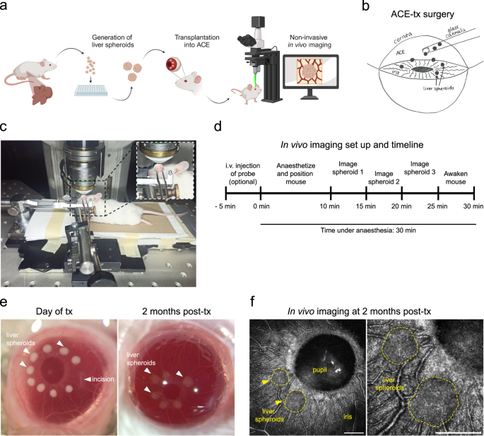
Longitudinal monitoring of liver function in vivo is hindered by the lack of high-resolution non-invasive imaging techniques. Using the anterior chamber of the mouse eye as a transplantation site, we have established a platform for longitudinal in vivo imaging of liver spheroids at cellular resolution. Transplanted liver spheroids engraft on the iris, become vascularized and innervated, retain hepatocyte-specific and liver-like features and can be studied by in vivo confocal microscopy. Employing fluorescent probes administered intravenously or spheroids formed from reporter mice, we showcase the potential use of this platform for monitoring hepatocyte cell cycle activity, bile secretion and lipoprotein uptake. Moreover, we show that hepatic lipid accumulation during diet-induced hepatosteatosis is mirrored in intraocular in vivo grafts. Here, we show a new technology which provides a crucial and unique tool to study liver physiology and disease progression in pre-clinical and basic research.


