2024-07-30 ペンシルベニア州立大学(PennState)
<関連情報>
- https://www.psu.edu/news/research/story/new-high-resolution-3d-maps-show-how-brains-blood-vessels-changes-age/
- https://www.nature.com/articles/s41467-024-50559-8
加齢がマウスの脳における脳血管網のリモデリングと機能的変化を引き起こす Aging drives cerebrovascular network remodeling and functional changes in the mouse brain
Hannah C. Bennett,Qingguang Zhang,Yuan-ting Wu,Steffy B. Manjila,Uree Chon,Donghui Shin,Daniel J. Vanselow,Hyun-Jae Pi,Patrick J. Drew & Yongsoo Kim
Nature Communications Published:30 July 2024
DOI:https://doi.org/10.1038/s41467-024-50559-8

Abstract
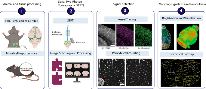
Aging is frequently associated with compromised cerebrovasculature and pericytes. However, we do not know how normal aging differentially impacts vascular structure and function in different brain areas. Here we utilize mesoscale microscopy methods and in vivo imaging to determine detailed changes in aged murine cerebrovascular networks. Whole-brain vascular tracing shows an overall ~10% decrease in vascular length and branching density with ~7% increase in vascular radii in aged brains. Light sheet imaging with 3D immunolabeling reveals increased arteriole tortuosity of aged brains. Notably, vasculature and pericyte densities show selective and significant reductions in the deep cortical layers, hippocampal network, and basal forebrain areas. We find increased blood extravasation, implying compromised blood-brain barrier function in aged brains. Moreover, in vivo imaging in awake mice demonstrates reduced baseline and on-demand blood oxygenation despite relatively intact neurovascular coupling. Collectively, we uncover regional vulnerabilities of cerebrovascular network and physiological changes that can mediate cognitive decline in normal aging.


