脳卒中による脳損傷に関する数十年来の説に再考を促す新たなデータ New data prompts reconsideration of decades-old theory about brain injury due to stroke
2022-04-21 ワシントン大学セントルイス

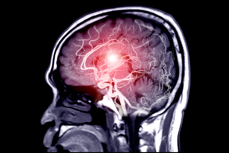
By scanning the genomes of nearly 6,000 stroke patients, researchers at Washington University School of Medicine in St. Louis identified two genes associated with recovery. Both are involved in regulating neuronal excitability, suggesting that targeting overstimulated neurons may help promote recovery in the pivotal first 24 hours. (Image: Getty Images)
脳卒中の後遺症として、過剰に興奮した神経細胞を落ち着かせることで、酸素不足ですでに損傷している神経細胞を殺す可能性のある毒性分子が放出されるのを防ぐことができると、神経科学者たちは考えていたのである。この考えは、細胞や動物を使った研究によって裏付けられていたが、多くの臨床試験で脳卒中患者の予後を改善できなかったため、2000年代前半には支持されなくなった。
しかし、新たなアプローチにより、この考えはあまりにも早急に捨て去られた可能性があることを示す証拠が得られた。この新しい知見は、『Brain』誌のオンライン版に掲載されている。
ワシントン大学医学部(セントルイス)の研究者らは、脳卒中を経験した約6,000人の全ゲノムをスキャンし、脳卒中後の極めて重要な最初の24時間以内の回復に関連する2つの遺伝子を特定しました。脳卒中の発症から24時間以内に起こる事象(良い事象も悪い事象も含む)は、脳卒中患者の長期的な回復への道筋をつけるものである。この2つの遺伝子は、いずれも神経細胞の興奮性の制御に関与していることが判明し、神経細胞の過剰な刺激が脳卒中の転帰に影響を及ぼすことを示す証拠となった。
<関連情報>
- https://source.wustl.edu/2022/04/calming-overexcited-neurons-may-protect-brain-after-stroke/
- https://academic.oup.com/brain/advance-article-abstract/doi/10.1093/brain/awac080/6537118?redirectedFrom=fulltext&login=false
多施設共同GWASにより、虚血性脳卒中後の転帰に興奮毒性が関連することが判明 Multi-ancestry GWAS reveals excitotoxicity associated with outcome after ischaemic stroke
Laura Ibanez, Laura Heitsch, Caty Carrera, Fabiana H. G. Farias, Jorge L. Del Aguila, Rajat Dhar, John Budde, Kristy Bergmann, Joseph Bradley, Oscar Harari,Chia-Ling Phuah, Robin Lemmens, Alessandro A. Viana Oliveira Souza, Francisco Moniche, Antonio Cabezas-Juan, Juan Francisco Arenillas, Jerzy Krupinksi, Natalia Cullell, Nuria Torres-Aguila, Elena Muiño, Jara Cárcel-Márquez, Joan Marti-Fabregas, Raquel Delgado-Mederos, Rebeca Marin-Bueno, Alejandro Hornick, Cristofol Vives-Bauza, Rosa Diaz Navarro, Silvia Tur, Carmen Jimenez, Victor Obach, Tomas Segura, Gemma Serrano-Heras, Jong-Won Chung, Jaume Roquer, Carol Soriano-Tarraga, Eva Giralt-Steinhauer, Marina Mola-Caminal, Joanna Pera, Katarzyna Lapicka-Bodzioch, Justyna Derbisz, Antoni Davalos, Elena Lopez-Cancio, Lucia Muñoz, Turgut Tatlisumak, Carlos Molina, Marc Ribo, Alejandro Bustamante, Tomas Sobrino, Jose Castillo-Sanchez, Francisco Campos, Emilio Rodriguez-Castro, Susana Arias-Rivas, Manuel Rodríguez-Yáñez, Christina Herbosa, Andria L. Ford, Alonso Gutierrez-Romero, Rodrigo Uribe-Pacheco, Antonio Arauz, Iscia Lopes-Cendes, Theodore Lowenkopf, Miguel A. Barboza, Hajar Amini, Boryana Stamova, Bradley P. Ander, Frank R Sharp, Gyeong Moon Kim, Oh Young Bang, Jordi Jimenez-Conde, Agnieszka Slowik, Daniel Stribian, Ellen A. Tsai, Linda C. Burkly, Joan Montaner, Israel Fernandez-Cadenas, Jin-Moo Lee, Carlos Cruchaga
Brain Published:25 February 2022
DOI:https://doi.org/10.1093/brain/awac080
Abstract
During the first hours after stroke onset neurological deficits can be highly unstable: some patients rapidly improve, while others deteriorate. This early neurological instability has a major impact on long-term outcome. Here, we aimed to determine the genetic architecture of early neurological instability measured by the difference between NIH stroke scale (NIHSS) within six hours of stroke onset and NIHSS at 24 h (ΔNIHSS). A total of 5,876 individuals from seven countries (Spain, Finland, Poland, United States, Costa Rica, Mexico and Korea) were studied using a multi-ancestry meta-analyses. We found that 8.7% of ΔNIHSS variance was explained by common genetic variations, and also that early neurological instability has a different genetic architecture than that of stroke risk. Eight loci (1p21.1, 1q42.2, 2p25.1, 2q31.2, 2q33.3, 5q33.2, 7p21.2,and 13q31.1) were genome-wide significant and explained 1.8% of the variability suggesting that additional variants influence early change in neurological deficits. We used functional genomics and bioinformatic annotation to identify the genes driving the association from each loci. eQTL mapping and SMR indicate that ADAM23 (log Bayes Factor (LBF) = 5.41) was driving the association for 2q33.3. Gene based analyses suggested that GRIA1 (LBF = 5.19), which is predominantly expressed in brain, is the gene driving the association for the 5q33.2 locus. These analyses also nominated GNPAT (LBF = 7.64)ABCB5 (LBF = 5.97) for the 1p21.1 and 7p21.1 loci. Human brain single nuclei RNA-seq indicates that the gene expression of ADAM23 and GRIA1 is enriched in neurons. ADAM23, a pre-synaptic protein, and GRIA1, a protein subunit of the AMPA receptor, are part of a synaptic protein complex that modulates neuronal excitability. These data provides the first genetic evidence in humans that excitotoxicity may contribute to early neurological instability after acute ischemic stroke.


