X線の着弾位置や線量を正確に検出することができれば、放射線治療による副次的な被害を軽減できる可能性がある The ability to accurately detect where X-rays land and in what dose could reduce the collateral damage from radiation therapy
2023-01-03 ミシガン大学
X線が体内の組織を熱したときに発生する小さな音波を捉えて増幅することで、医療専門家は体内の放射線量をマッピングし、リアルタイムで治療の指針となる新しいデータを得ることができるのです。これは、これまで医師が「見る」ことができなかった相互作用を見ることができる、世界初の試みです。
Jonathan Rubin Collegiate Professor of Biomedical Engineering、放射線学教授で、Nature Biotechnology誌の研究のcorresponding authorであるXueding Wangは述べています。彼はまた、U-Mの光学イメージング研究所を率いています。
放射線治療は、腫瘍周辺の健康な細胞を殺したり傷つけたりすることが多いため、精度の低さがこの利点を損なっている。また、新たながんが発生するリスクも高くなります。
リアルタイムの3D画像を使えば、医師は放射線をより正確にがん細胞に向けて照射し、隣接する組織の被ばくを抑えることができます。そのためには、単に “聞く “ことが必要なのです。
X線が体内の組織に吸収されると、熱エネルギーに変換されます。その熱によって組織は急速に膨張し、その膨張によって音波が発生する。
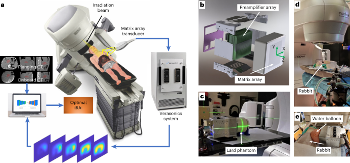
この音波は微弱で、一般的な超音波診断技術では通常検出できない。U-Mの新しい電離放射線音響画像システムは、患者側に配置された超音波トランスデューサーの配列で音波を検出する。信号は増幅され、画像再構成のために超音波装置に転送される。
この画像をもとに、腫瘍クリニックでは、より安全で効果的な治療を行うために、治療中の放射線のレベルや軌道を変更することができます。
「将来的には、放射線治療中の位置や臓器の動き、解剖学的な変化から生じる不確実性を画像情報で補正できるようになるでしょう」と、生体医工学研究員でこの研究の筆頭著者であるWei Zhangは述べています。「と、この研究の筆頭著者である生体工学の研究者Wei Zhangは言いました。「そうすれば、ピンポイントで癌腫瘍に線量を供給することができます。
U-Mの技術のもう一つの利点は、臨床医が慣れているプロセスを劇的に変えることなく、現在の放射線治療機器に簡単に追加できることです。
<関連情報>
- https://news.umich.edu/tracking-radiation-treatment-in-real-time-promises-safer-more-effective-cancer-therapy/
- https://www.nature.com/articles/s41587-022-01593-8
がん治療中の肝臓深部への放射線量到達のリアルタイムボリュームイメージング Real-time, volumetric imaging of radiation dose delivery deep into the liver during cancer treatment
Wei Zhang,Ibrahim Oraiqat,Dale Litzenberg,Kai-Wei Chang,Scott Hadley,Noora Ba Sunbul,Martha M. Matuszak,Christopher J. Tichacek,Eduardo G. Moros,Paul L. Carson,Kyle C. Cuneo,Xueding Wang & Issam El Naqa
Nature Biotechnology Published:02 January 2023
DOI:https://doi.org/10.1038/s41587-022-01593-8

Abstract
Ionizing radiation acoustic imaging (iRAI) allows online monitoring of radiation’s interactions with tissues during radiation therapy, providing real-time, adaptive feedback for cancer treatments. We describe an iRAI volumetric imaging system that enables mapping of the three-dimensional (3D) radiation dose distribution in a complex clinical radiotherapy treatment. The method relies on a two-dimensional matrix array transducer and a matching multi-channel preamplifier board. The feasibility of imaging temporal 3D dose accumulation was first validated in a tissue-mimicking phantom. Next, semiquantitative iRAI relative dose measurements were verified in vivo in a rabbit model. Finally, real-time visualization of the 3D radiation dose delivered to a patient with liver metastases was accomplished with a clinical linear accelerator. These studies demonstrate the potential of iRAI to monitor and quantify the 3D radiation dose deposition during treatment, potentially improving radiotherapy treatment efficacy using real-time adaptive treatment.


