2025-06-05 神戸大学

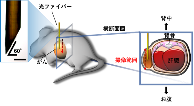
図1. 本研究で開発した顕微内視鏡イメージングシステムの概略図
左上の写真は、内視鏡プローブとして用いる光ファイバーを示している。スケールバー=0.2 mm。内視鏡プローブを、がんの表面から刺入し、撮像しながら一定速度で反対側の端まで進めることで、がんの端から端まで撮像することが可能である。
<関連情報>
- https://www.kobe-u.ac.jp/ja/news/article/20250605-66691/
- https://www.cell.com/cell-reports-methods/fulltext/S2667-2375(25)00092-X
微小内視鏡によるがん細胞のエンド・ツー・エンド定期的腫瘍イメージング Microendoscopy for periodic intravital end-to-end tumor imaging of cancer cells
Toshiyuki Goto ∙ Masayuki Nakano ∙ Sally Danno ∙ … ∙ Kei Mizuno ∙ Yosky Kataoka ∙ Kazuo Funabiki
Cell Reports methods Published:June 4, 2025
DOI:https://doi.org/10.1016/j.crmeth.2025.101056
Motivation
The heterogeneity in cancer cell behaviors in tumor is considered the main reason for the difficulty in achieving complete remission. To determine the efficacy of anticancer treatments, the behavior of cancer cells should be investigated at a cellular resolution in all the different micro- and macroenvironments within each tumor. However, current intravital imaging methods do not realize that. Thus, we developed an optical-fiber-bundle-based microendoscopy with a fluorescent ubiquitination-based cell- cycle indicator (Fucci) system to achieve the intravital, periodic, and multicolor end-to-end tumor imaging of the proliferative activity of cancer cells at a cellular-level resolution.
Highlights
- We present a microendoscopy method for intravital imaging of cancer cells
- We demonstrate live imaging of Fucci-reporter cancer cells in tumor-bearing mice
- Proliferation and nuclear enlargement of the cells are spatiotemporally visualized
- We report complex temporal changes in these cell states in response to anticancer drugs
Summary
The spatiotemporal heterogeneity in intratumor proliferative behavior of cancer cells deeply affects tumor environment characteristics and the efficacy of anticancer treatments. Thus, intravital imaging with unlimited imaging depth and cellular-level resolution is greatly desired. We developed an optical-fiber-bundle-based microendoscope with a genetically encoded fluorescent ubiquitination-based cell-cycle indicator (Fucci) system to achieve the intravital, periodic, and multicolor end-to-end imaging of the proliferative activity of cancer cells at a cellular-level resolution. This technique enabled the periodic visualization of spatiotemporal cellular responses, including cell-cycle arrest and resumption, and nuclear enlargement following the administration of anticancer drugs in living mice. It was suggested that proliferating cell ratio and nuclear enlargement in cancer cells at the surface region of tumor characterized by abundant vascular invasion contribute to aggressive tumor regrowth after chemotherapy. The application of this technique can accelerate innovation in cancer biology and therapeutics.

