2024-04-24 カリフォルニア大学サンディエゴ校(UCSD)
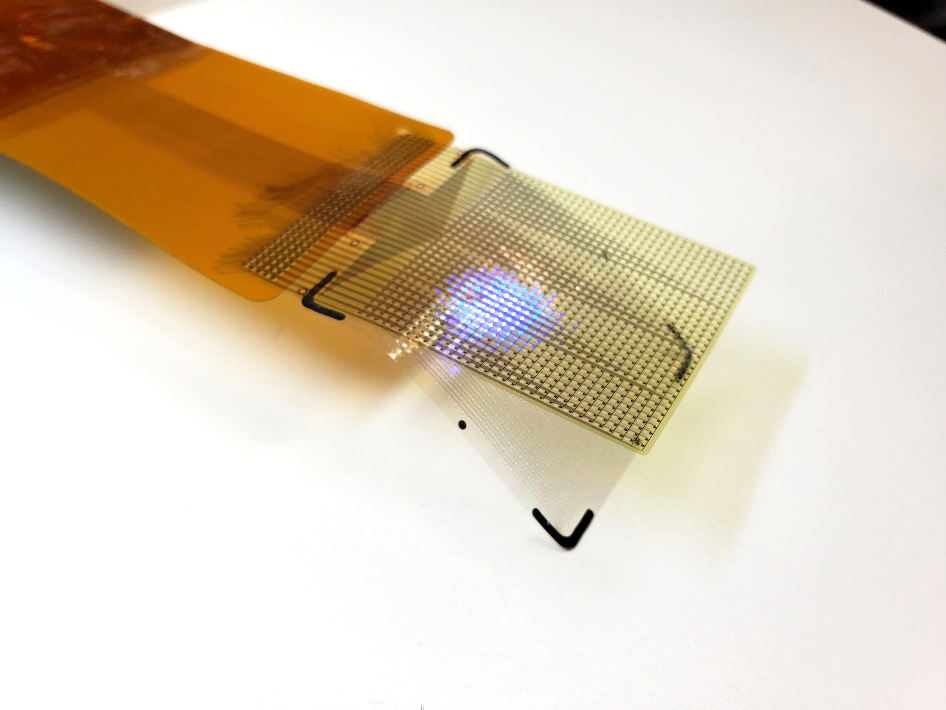
 The device’s LEDs can light up in several colors. This allows surgeons to see which areas they need to operate on. It allows them to track brain states during surgery, including the onset of epileptic seizures.
<関連情報>
- https://today.ucsd.edu/story/a-flexible-microdisplay-can-monitor-and-visualize-brain-activity-in-real-time-during-brain-surgery
- https://www.science.org/doi/10.1126/scitranslmed.adj7257
- https://www.science.org/doi/10.1126/scitranslmed.abj1441
脳表面の神経細胞活動を可視化する脳波マイクロディスプレイ An electroencephalogram microdisplay to visualize neuronal activity on the brain surface
YOUNGBIN TCHOE, TIANHAI WU, HOI SANG U, DAVID M. ROTH, […], AND SHADI A. DAYEH
Science Translational Medicine Published:24 Apr 2024
DOI:https://doi.org/10.1126/scitranslmed.adj7257
Editor’s summary
The visualization of recorded cortical activity in real time on the brain surface could facilitate electrophysiological studies and neurosurgical procedures. Here, Tchoe et al. combined micro-electrocorticography electrode grids with light-emitting diodes to create intracranial electroencephalography microdisplays. The authors show in a series of proof-of-concept experiments in pigs and rats that these microdisplays allow the visualization of spontaneous or elicited cortical activity and functional cortical boundaries on the surface of the brain. Future iterations of this system could also be useful in clinical settings. —Daniela Neuhofer
Abstract
Functional mapping during brain surgery is applied to define brain areas that control critical functions and cannot be removed. Currently, these procedures rely on verbal interactions between the neurosurgeon and electrophysiologist, which can be time-consuming. In addition, the electrode grids that are used to measure brain activity and to identify the boundaries of pathological versus functional brain regions have low resolution and limited conformity to the brain surface. Here, we present the development of an intracranial electroencephalogram (iEEG)–microdisplay that consists of freestanding arrays of 2048 GaN light-emitting diodes laminated on the back of micro-electrocorticography electrode grids. With a series of proof-of-concept experiments in rats and pigs, we demonstrate that these iEEG-microdisplays allowed us to perform real-time iEEG recordings and display cortical activities by spatially corresponding light patterns on the surface of the brain in the surgical field. Furthermore, iEEG-microdisplays allowed us to identify and display cortical landmarks and pathological activities from rat and pig models. Using a dual-color iEEG-microdisplay, we demonstrated coregistration of the functional cortical boundaries with one color and displayed the evolution of electrical potentials associated with epileptiform activity with another color. The iEEG-microdisplay holds promise to facilitate monitoring of pathological brain activity in clinical settings.
多チャンネルPtNRGridsによるヒト脳マッピングは時空間ダイナミクスを解像する Human brain mapping with multithousand-channel PtNRGrids resolves spatiotemporal dynamics
YOUNGBIN TCHOE, ANDREW M. BOURHIS, DANIEL R. CLEARY, BRITTANY STEDELIN, […], AND SHADI A. DAYEH
Science Translational Medicine Published:19 Jan 2022
DOI:https://doi.org/10.1126/scitranslmed.abj1441

Cortex in high resolution
Recording brain cortical activity with high spatial and temporal resolution is critical for understanding brain circuitry in physiological and pathological conditions. In this study, Tchoe et al. developed a reconfigurable and scalable thin-film, multithousand-channel neurophysiological recording grids using platinum nanorods, called PtNRGrids, that could record thousands of channels with submillimeter resolution in the rat barrel cortex. In human subjects, PtNRGrids were able to provide high-resolution recordings of large and curvilinear brain areas and to resolve spatiotemporal dynamics of motor and sensory activities. The results suggest that PtNRGrids could be used in the preclinical and clinical setting for high spatial and temporal recording of neural activity.
Abstract
Electrophysiological devices are critical for mapping eloquent and diseased brain regions and for therapeutic neuromodulation in clinical settings and are extensively used for research in brain-machine interfaces. However, the existing clinical and experimental devices are often limited in either spatial resolution or cortical coverage. Here, we developed scalable manufacturing processes with a dense electrical connection scheme to achieve reconfigurable thin-film, multithousand-channel neurophysiological recording grids using platinum nanorods (PtNRGrids). With PtNRGrids, we have achieved a multithousand-channel array of small (30 μm) contacts with low impedance, providing high spatial and temporal resolution over a large cortical area. We demonstrated that PtNRGrids can resolve submillimeter functional organization of the barrel cortex in anesthetized rats that captured the tissue structure. In the clinical setting, PtNRGrids resolved fine, complex temporal dynamics from the cortical surface in an awake human patient performing grasping tasks. In addition, the PtNRGrids identified the spatial spread and dynamics of epileptic discharges in a patient undergoing epilepsy surgery at 1-mm spatial resolution, including activity induced by direct electrical stimulation. Collectively, these findings demonstrated the power of the PtNRGrids to transform clinical mapping and research with brain-machine interfaces.


