2023-04-19 マサチューセッツ工科大学(MIT)

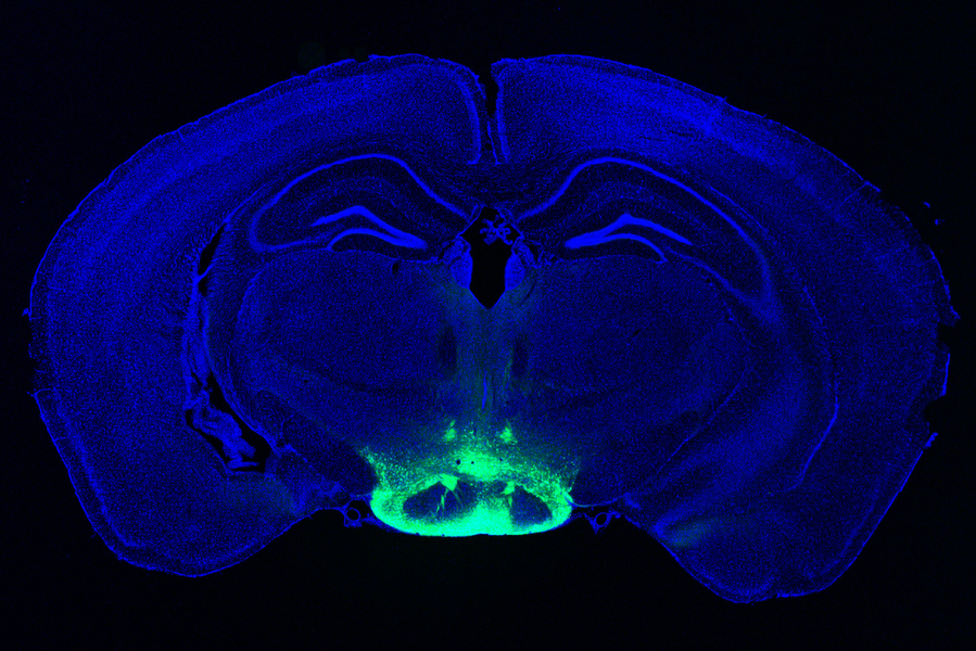
MIT researchers have identified neurons in the brain that may contribute to some of the earliest symptoms of Alzheimer’s disease, making it a good target for potential new drugs to treat the disease. The area these neurons are found, in a part of the hypothalamus called the mammillary body, is highlighted in green.
Image: Courtesy of the researchers
研究グループは、てんかんの治療に用いられる薬剤で治療することで、記憶障害を回復させることができることを発見しました。
現在、乳頭体の外側ニューロンが脳の他の部位とどのようにつながっているのかを定義し、記憶回路を形成する仕組みを解明することに取り組んでいるという。
<関連情報>
- https://news.mit.edu/2023/neuroscientists-identify-cells-vulnerable-alzheimers-0419
- https://www.science.org/doi/10.1126/scitranslmed.abq1019
マウス脳の外側乳頭体神経細胞はアルツハイマー病で不釣り合いなほど傷つきやすい Lateral mammillary body neurons in mouse brain are disproportionately vulnerable in Alzheimer’s disease
Wen-Chin Huang,Zhuyu Peng,Mitchell H. Murdock,Liwang Liu,Hansruedi Mathys,Jose Davila-Velderrain,Xueqiao Jiang,Maggie Chen,Ayesha P. Ng,TaeHyun Kim,Fatema Abdurrob,Fan Gao,David A. Bennett,Manolis Kellis and Li-Huei Tsai
Science Translational Medicine Published:19 Apr 2023
DOI:https://doi.org/10.1126/scitranslmed.abq1019
Abstract
The neural circuits governing the induction and progression of neurodegeneration and memory impairment in Alzheimer’s disease (AD) are incompletely understood. The mammillary body (MB), a subcortical node of the medial limbic circuit, is one of the first brain regions to exhibit amyloid deposition in the 5xFAD mouse model of AD. Amyloid burden in the MB correlates with pathological diagnosis of AD in human postmortem brain tissue. Whether and how MB neuronal circuitry contributes to neurodegeneration and memory deficits in AD are unknown. Using 5xFAD mice and postmortem MB samples from individuals with varying degrees of AD pathology, we identified two neuronal cell types in the MB harboring distinct electrophysiological properties and long-range projections: lateral neurons and medial neurons. lateral MB neurons harbored aberrant hyperactivity and exhibited early neurodegeneration in 5xFAD mice compared with lateral MB neurons in wild-type littermates. Inducing hyperactivity in lateral MB neurons in wild-type mice impaired performance on memory tasks, whereas attenuating aberrant hyperactivity in lateral MB neurons ameliorated memory deficits in 5xFAD mice. Our findings suggest that neurodegeneration may be a result of genetically distinct, projection-specific cellular dysfunction and that dysregulated lateral MB neurons may be causally linked to memory deficits in AD.
A vulnerable neuronal subtype in Alzheimer’s disease
How brain neural circuit dysfunction, pathological insults, and the demise of neurons drive memory deficits in Alzheimer’s disease (AD) remains poorly understood. The mammillary body, a subregion of the hypothalamus, exhibits early amyloid deposition in the 5xFAD mouse model of AD. In a new study, Huang et al. used single-cell RNA sequencing of postmortem brain sections of mouse mammillary body and brain slice electrophysiology to show that lateral mammillary body neurons of 5xFAD mice harbored aberrant hyperactivity that correlated with impaired memory in two behavioral tests compared with wild-type littermate control animals. These findings implicate this neuronal subtype in the pathogenesis of AD. —OS

