2023-12-13 カリフォルニア大学バークレー校(UCB)
◆視覚情報を処理し、それを脳の他の部分に伝達する多様な細胞タイプを含む視網膜は、多くの細胞タイプが古代の進化の歴史を持っているという研究結果から、哺乳動物の最後の共通の祖先の視網膜は約2億年前に現代の哺乳動物の視網膜と同等の複雑さを持っていた可能性があることが示唆されています。これにより、ヒトの眼病の研究に役立つであろう新しい細胞タイプの地図が得られました。
<関連情報>
- https://news.berkeley.edu/2023/12/13/cell-types-in-the-eye-have-ancient-evolutionary-origins
- https://www.nature.com/articles/s41586-023-06638-9
- https://www.nature.com/articles/s41586-023-06659-4
- https://www.sciencedirect.com/science/article/pii/S0092867419300376
脊椎動物網膜における神経細胞クラスとタイプの進化 Evolution of neuronal cell classes and types in the vertebrate retina
Joshua Hahn,Aboozar Monavarfeshani,Mu Qiao,Allison H. Kao,Yvonne Kölsch,Ayush Kumar,Vincent P. Kunze,Ashley M. Rasys,Rose Richardson,Joseph B. Wekselblatt,Herwig Baier,Robert J. Lucas,Wei Li,Markus Meister,Joshua T. Trachtenberg,Wenjun Yan,Yi-Rong Peng,Joshua R. Sanes & Karthik Shekhar
Nature Published:13 December 2023
DOI:https://doi.org/10.1038/s41586-023-06638-9
")
Abstract
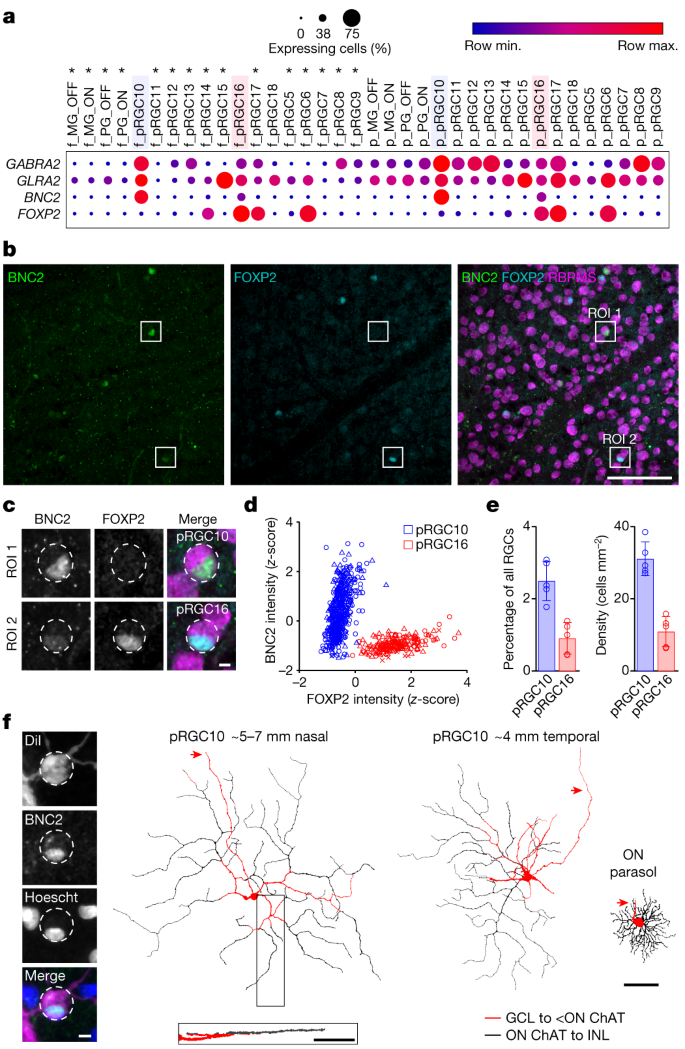
The basic plan of the retina is conserved across vertebrates, yet species differ profoundly in their visual needs1. Retinal cell types may have evolved to accommodate these varied needs, but this has not been systematically studied. Here we generated and integrated single-cell transcriptomic atlases of the retina from 17 species: humans, two non-human primates, four rodents, three ungulates, opossum, ferret, tree shrew, a bird, a reptile, a teleost fish and a lamprey. We found high molecular conservation of the six retinal cell classes (photoreceptors, horizontal cells, bipolar cells, amacrine cells, retinal ganglion cells (RGCs) and Müller glia), with transcriptomic variation across species related to evolutionary distance. Major subclasses were also conserved, whereas variation among cell types within classes or subclasses was more pronounced. However, an integrative analysis revealed that numerous cell types are shared across species, based on conserved gene expression programmes that are likely to trace back to an early ancestral vertebrate. The degree of variation among cell types increased from the outer retina (photoreceptors) to the inner retina (RGCs), suggesting that evolution acts preferentially to shape the retinal output. Finally, we identified rodent orthologues of midget RGCs, which comprise more than 80% of RGCs in the human retina, subserve high-acuity vision, and were previously believed to be restricted to primates2. By contrast, the mouse orthologues have large receptive fields and comprise around 2% of mouse RGCs. Projections of both primate and mouse orthologous types are overrepresented in the thalamus, which supplies the primary visual cortex. We suggest that midget RGCs are not primate innovations, but are descendants of evolutionarily ancient types that decreased in size and increased in number as primates evolved, thereby facilitating high visual acuity and increased cortical processing of visual information.
霊長類網膜におけるON型方向選択性神経節細胞 An ON-type direction-selective ganglion cell in primate retina
Anna Y. M. Wang,Manoj M. Kulkarni,Amanda J. McLaughlin,Jacqueline Gayet,Benjamin E. Smith,Max Hauptschein,Cyrus F. McHugh,Yvette Y. Yao & Teresa Puthussery
Nature Published:25 October 2023
DOI:https://doi.org/10.1038/s41586-023-06659-4
Abstract
To maintain a stable and clear image of the world, our eyes reflexively follow the direction in which a visual scene is moving. Such gaze-stabilization mechanisms reduce image blur as we move in the environment. In non-primate mammals, this behaviour is initiated by retinal output neurons called ON-type direction-selective ganglion cells (ON-DSGCs), which detect the direction of image motion and transmit signals to brainstem nuclei that drive compensatory eye movements1. However, ON-DSGCs have not yet been identified in the retina of primates, raising the possibility that this reflex is mediated by cortical visual areas. Here we mined single-cell RNA transcriptomic data from primate retina to identify a candidate ON-DSGC. We then combined two-photon calcium imaging, molecular identification and morphological analysis to reveal a population of ON-DSGCs in the macaque retina. The morphology, molecular signature and GABA (γ-aminobutyric acid)-dependent mechanisms that underlie direction selectivity in primate ON-DSGCs are highly conserved with those in other mammals. We further identify a candidate ON-DSGC in human retina. The presence of ON-DSGCs in primates highlights the need to examine the contribution of subcortical retinal mechanisms to normal and aberrant gaze stabilization in the developing and mature visual system.
霊長類網膜における眼窩細胞と周辺細胞の分子分類と比較分類学 Molecular Classification and Comparative Taxonomics of Foveal and Peripheral Cells in Primate Retina
Yi-Rong Peng, Karthik Shekhar, Wenjun Yan, Dustin Herrmann, Anna Sappington, Gregory S. Bryman, Tavé van Zyl, Michael Tri. H. Do, Aviv Regev, Joshua R. Sanes
Cell Published: January 31, 2019
DOI:https://doi.org/10.1016/j.cell.2019.01.004
Highlights
•Macaque fovea and peripheral retina each contain >65 cell types
•Most types correspond between regions but differ in proportions and gene expression
•Greater conservation of interneuron than ganglion cell types between macaque and mouse
•Cell-type- and region-specific expression of genes implicated in human blindness
Summary
High-acuity vision in primates, including humans, is mediated by a small central retinal region called the fovea. As more accessible organisms lack a fovea, its specialized function and its dysfunction in ocular diseases remain poorly understood. We used 165,000 single-cell RNA-seq profiles to generate comprehensive cellular taxonomies of macaque fovea and peripheral retina. More than 80% of >60 cell types match between the two regions but exhibit substantial differences in proportions and gene expression, some of which we relate to functional differences. Comparison of macaque retinal types with those of mice reveals that interneuron types are tightly conserved. In contrast, projection neuron types and programs diverge, despite exhibiting conserved transcription factor codes. Key macaque types are conserved in humans, allowing mapping of cell-type and region-specific expression of >190 genes associated with 7 human retinal diseases. Our work provides a framework for comparative single-cell analysis across tissue regions and species.
Graphical Abstract

")
