2026-11-05 清華大学
<関連情報>
- https://www.tsinghua.edu.cn/en/info/1245/15110.htm
- https://link.springer.com/article/10.1186/s12916-026-05256-2
3Dバイオプリントされたヒト血管オルガノイドシートは、機能的な虚血修復を促進し、生体内での適応的なリモデリングを示す 3D bioprinted human vascular organoid sheets promote functional ischemic repair and exhibit adaptive in vivo remodeling
Xinyu Fu, Li Yan, Zhaosen Chen, Bohan Dou, Dezhi Zhou, Xinyao Zhou, Kengyuan Qu, Che Gao, Peiliang Wang, Fengzhi Zhang, Zihao Zou, Taoxia Wang, Guiying Li, Liliang Ouyang & Jie Na
BMC Medicine Published:25 September 2026
DOI:https://doi.org/10.1186/s12916-026-05256-2
Abstract
Background
Ischemic vascular diseases remain a major clinical challenge, creating a need for engineered vascular tissues that can establish functional vascular networks and promote durable tissue repair. Conventional vascular organoids offer limited control over cellular composition and spatial organization, while the fate and adaptive remodeling of graft-derived human vascular cells after transplantation remain poorly understood.
Methods
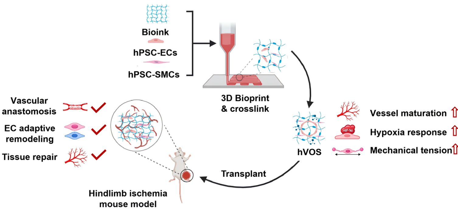
Human vascular organoid sheets (hVOS) were constructed by extrusion-based three-dimensional (3D) bioprinting of human pluripotent stem cell (hPSC)-derived endothelial cells (ECs) and smooth muscle cells (SMCs) at a defined ratio within a gelatin methacryloyl (GelMA)-based bioink under chemically defined conditions. Vascular organization and cellular states were characterized using functional assays, immunofluorescence imaging, and single-cell RNA sequencing (scRNA-seq). Therapeutic efficacy and graft remodeling were evaluated in a murine hindlimb ischemia model using laser speckle perfusion imaging, histological analysis, intravital two-photon imaging, and scRNA-seq of recovered graft-derived human cells.
Results
Co-bioprinting ECs and SMCs accelerated vascular network formation and generated stable, interconnected vascular structures that underwent progressive maturation during culture. scRNA-seq identified diverse vascular and stromal populations and revealed transcriptional programs associated with vascular maturation, mechanotransduction, and hypoxic adaptation. Following transplantation, hVOS significantly improved blood perfusion, increased limb salvage, and promoted ischemic tissue repair. Intravital imaging detected circulating dextran within GFP-labeled hVOS-derived vascular structures at days 14 and 28, demonstrating perfusion of graft-derived vascular structures by the host circulation. Post-transplantation scRNA-seq revealed substantial adaptive remodeling of graft-derived ECs toward venous-biased and inflammatory states, accompanied by activation of NF-κB- and stress-associated programs. Transplanted SMCs and fibroblasts also exhibited coordinated transcriptional changes associated with wound healing and extracellular matrix remodeling.
Conclusions
hVOS provide a reproducible and design-flexible 3D-bioprinted vascular tissue platform that enables controlled multicellular organization and formation of prevascularized constructs while supporting vascular integration and ischemic tissue repair in vivo. Single-cell analyses further reveal substantial adaptive remodeling of graft-derived vascular and stromal cells following transplantation. These findings support hVOS as a versatile platform for vascular regenerative medicine and for investigating the in vivo behavior of engineered human vascular tissues.