マウスの様々な脳領域や細胞型が血管系に支えられていることを示す新しい地図が完成。 New maps help researchers study how various mouse brain regions, cell types are supported by vascular system
2022-07-07 ペンシルベニア州立大学(PennState)
研究チームは、マウスのさまざまな脳領域で血管細胞や構造がどのように異なるかを示す新しいマップを作成し、血管供給が神経疾患にどのように関係しているかを理解するのに役立てたいと考えている。
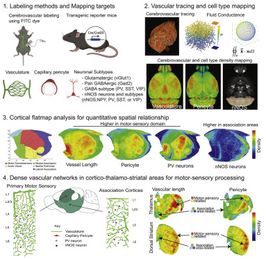
研究チームは、運動や感覚情報を処理する脳の領域には、血管系が多く存在することを発見した。また、異なる情報を合成して処理する脳の連合領域は、血管の程度が低くなる傾向があることも分かりました。
研究チームは、加齢に伴う神経細胞の減少は、脳内のエネルギー基盤の機能不全の結果ではないかとの仮説を立てている。
<関連情報>
- https://www.psu.edu/news/research/story/brain-regions-vulnerable-disease-may-lack-adequate-energy-blood-supply/
- https://www.cell.com/cell-reports/fulltext/S2211-1247(22)00764-1
マウスにおける脳血管網と神経細胞型の定量的関係 Quantitative relationship between cerebrovascular network and neuronal cell types in mice
Yuan-ting Wu ,Hannah C. Bennett ,Uree Chon,Daniel J. Vanselow,Qingguang Zhang,Rodrigo Muñoz-Castañeda,Keith C. Cheng ,Pavel Osten ,Patrick J. Drew ,Yongsoo Kim
Cell Reports Published:JUNE 21, 2022
DOI:https://doi.org/10.1016/j.celrep.2022.110978

Highlights
•Resource to examine cerebrovasculature, pericytes, and related neuronal subtypes
•Dense cerebrovasculature network in motor sensory circuits
•Cortical vasculature significantly correlated with parvalbumin and nNOS neurons
•Neurovascular architecture to meet regionally distinct brain energy regulation
Summary
The cerebrovasculature and its mural cells must meet brain regional energy demands, but how their spatial relationship with different neuronal cell types varies across the brain remains largely unknown. Here we apply brain-wide mapping methods to comprehensively define the quantitative relationships between the cerebrovasculature, capillary pericytes, and glutamatergic and GABAergic neurons, including neuronal nitric oxide synthase-positive (nNOS+) neurons and their subtypes in adult mice. Our results show high densities of vasculature with high fluid conductance and capillary pericytes in primary motor sensory cortices compared with association cortices that show significant positive and negative correlations with energy-demanding parvalbumin+ and vasomotor nNOS+ neurons, respectively. Thalamo-striatal areas that are connected to primary motor sensory cortices also show high densities of vasculature and pericytes, suggesting dense energy support for motor sensory processing areas. Our cellular-resolution resource offers opportunities to examine spatial relationships between the cerebrovascular network and neuronal cell composition in largely understudied subcortical areas.

