2024-06-04 マックス・プランク研究所
<関連情報>
- https://www.mpg.de/22021501/0603-vasb-20240604-bone-formation-in-skull-154090-x
- https://www.nature.com/articles/s41467-024-48579-5
腓骨再生における血管新生は骨新生から切り離される Angiogenesis is uncoupled from osteogenesis during calvarial bone regeneration
M. Gabriele Bixel,Kishor K. Sivaraj,Melanie Timmen,Vishal Mohanakrishnan,Anusha Aravamudhan,Susanne Adams,Bong-Ihn Koh,Hyun-Woo Jeong,Kai Kruse,Richard Stange & Ralf H. Adams
Nature Communications Published:04 June 2024
DOI:https://doi.org/10.1038/s41467-024-48579-5

Abstract
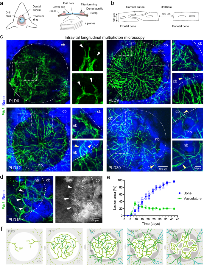
Bone regeneration requires a well-orchestrated cellular and molecular response including robust vascularization and recruitment of mesenchymal and osteogenic cells. In femoral fractures, angiogenesis and osteogenesis are closely coupled during the complex healing process. Here, we show with advanced longitudinal intravital multiphoton microscopy that early vascular sprouting is not directly coupled to osteoprogenitor invasion during calvarial bone regeneration. Early osteoprogenitors emerging from the periosteum give rise to bone-forming osteoblasts at the injured calvarial bone edge. Microvessels growing inside the lesions are not associated with osteoprogenitors. Subsequently, osteogenic cells collectively invade the vascularized and perfused lesion as a multicellular layer, thereby advancing regenerative ossification. Vascular sprouting and remodeling result in dynamic blood flow alterations to accommodate the growing bone. Single cell profiling of injured calvarial bones demonstrates mesenchymal stromal cell heterogeneity comparable to femoral fractures with increase in cell types promoting bone regeneration. Expression of angiogenesis and hypoxia-related genes are slightly elevated reflecting ossification of a vascularized lesion site. Endothelial Notch and VEGF signaling alter vascular growth in calvarial bone repair without affecting the ossification progress. Our findings may have clinical implications for bone regeneration and bioengineering approaches.


