
2026-08-17 コロンビア大学
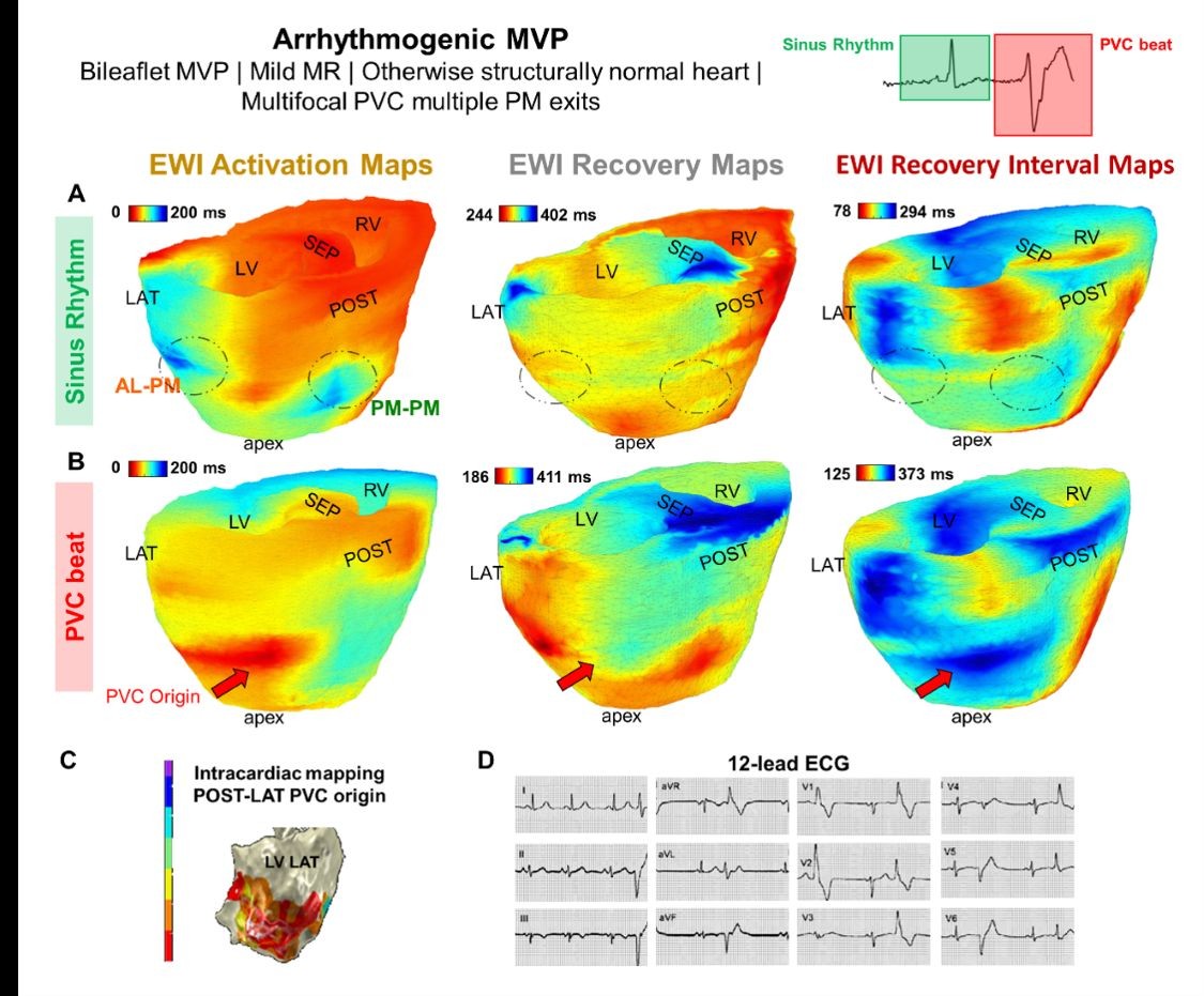
EWI imaging of a 22-year-old female patient with mitral valve prolapse and an abnormal heart rhythm.
<関連情報>
- https://www.engineering.columbia.edu/about/news/doctors-may-now-see-hidden-signs-sudden-cardiac-death
- https://www.pnas.org/doi/10.1073/pnas.2503559123
小児僧帽弁疾患の特徴付けのための電気機械的活性化および回復波イメージング Electromechanical activation and recovery wave imaging for pediatric mitral valve disease characterization
Melina Tourni, Christina Proestaki, Seungyeon Julia Han, +12 , and Elisa E. Konofagou
Proceedings of the National Academy of Sciences Published:August 17, 2026
DOI:https://doi.org/10.1073/pnas.2503559123
Abstract
Mitral valve (MV) disease, particularly MV prolapse (MVP) and mitral regurgitation (MR), affects 2 to 5% of the population and poses a substantial arrhythmogenic risk, with 43% of MVP patients developing arrhythmias. Although less common in children, the absence of comorbidities can uniquely isolate early electromechanical alterations that may elucidate mechanisms later contributing to arrhythmic risk and devastating effects such as sudden cardiac death. Conventional echocardiography lacks sensitivity for MV electromechanics, motivating advanced approaches. We introduce Electromechanical Wave Imaging (EWI), a high-frame-rate echocardiography modality, to map MV-complex activation and diastolic recovery in N = 21 MVP, MR, and control pediatric subjects (13.10 ± 4.51 y old, 43% male). A preclinical canine study (n = 3) established EWI’s ability to observe temporally coupled electromechanical wave propagation across the atrioventricular junction through the closed MV-following atrial and preceding ventricular activation (73.0 ms, 60 BPM). MVP patients exhibited significantly delayed left ventricular (LV) activation (76.04 ± 12.51 ms vs. 47.64 ± 2.57 ms in controls, P = 0.0013), primarily in papillary muscles. Both MVP and MR-only subjects exhibited prolonged LV recovery, with MR-only patients showing significantly longer recovery intervals (MR-only: 277.0 ± 27.61 ms, Control: 248.7 ± 10.43 ms, MVP vs. Control: MR-only vs. Control: P = 0.0395). In two arrhythmogenic MVP cases, EWI localized arrhythmic exit sites adjacent to LV papillary muscles, coinciding with regional delayed sinus activation, aligning with invasive electrophysiology. This study demonstrates that atrioventricular valve function dictates cardiac electromechanical function and establishes full-cycle EWI as a transformative tool for diagnosing MV disease electromechanical effects, assessing arrhythmic risk, guiding interventions, and advancing noninvasive cardiac imaging.

