2026-09-25 東北大学
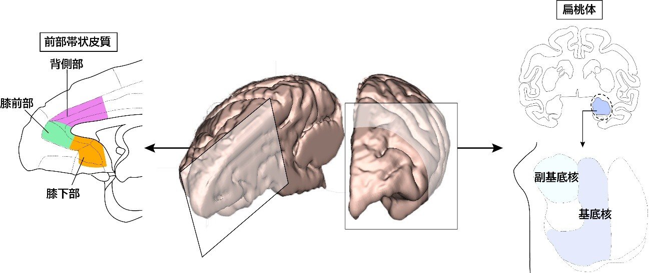
図1. サルの脳における前部帯状皮質と扁桃体の位置を模式的に示しています。前部帯状皮質は大脳の内側面で脳梁を取り囲むように位置し、機能的に膝前部、膝下部、背側部の3つの小領域に分けられます。扁桃体は側頭葉の内側の深部に位置しています。本研究では特に前部帯状皮質の小領域から扁桃体の基底核と副基底核への神経投射を解析しました。
<関連情報>
- https://www.tohoku.ac.jp/japanese/2026/09/press20260925-04-brain.html
- https://www.jneurosci.org/content/46/37/e0671262026
霊長類における前帯状皮質から扁桃体への投射:感情と気分調節の基盤となる地形的および層特異的な組織 Anterior Cingulate Cortex Projections to the Amygdala in Primates: Topographic and Layer-Specific Organization Underlying Emotion and Mood Regulation
Kei Kimura, Yuki Soga, Rintaro Yoshino, Andi Zheng, Satoshi Nonomura, Gaoge Yan, Soshi Tanabe, Shinya Nakamura, Shinya Ohara, Ken-ichi Inoue, Masahiko Takada and Ken-Ichiro Tsutsui
The Journal of Neuroscience Published:16 September 2026
Abstract
Emotion and mood regulation critically depends on interactions between the anterior cingulate cortex (ACC) and the amygdala. However, the detailed architecture of ACC projections to their major targets, the basal (BA) and accessory (AcBA) basal nuclei of the amygdala, remains unclear. To address this issue, a combined retrograde and anterograde tracing with viral vectors was performed in macaques of either sex to map the projection patterns from the pregenual (pgACC), subgenual (sgACC), and dorsal (dACC) subareas. Data revealed that ACC neurons projecting to the BA arose predominantly from the superficial layers (II/III) of all subareas and the deep layers (V/VI) of the sgACC, whereas ACC neurons projecting to the AcBA originated mainly in the deep layers of the sgACC and dACC. The present study defines the topographic and layer-specific organization of ACC–amygdala connectivity in primates and subserves to provide an anatomical basis for future causal and translational approaches, such as targeted interventions against ACC-related mood disorders.