2026-10-16 東京科学大学
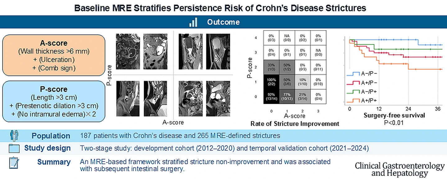
図1. 腸管狭窄に対する評価スコアを開発
<関連情報>
- https://www.isct.ac.jp/ja/news/nzh9dpo52x97
- https://www.cghjournal.org/article/S1542-3565(26)00681-6/fulltext
ベースライン磁気共鳴腸管造影検査によるクローン病狭窄の持続リスクの層別化 Baseline Magnetic Resonance Enterography Stratifies Persistence Risk of Crohn’s Disease Strictures
Kento Takenaka ∙ Junichi Tsuchiya ∙ Ami Kawamoto ∙ … ∙ Toshimitsu Fujii ∙ Kazuo Ohtsuka ∙ Ryuichi Okamoto
Clinical Gastroenterology and Hepatology Published:September 15, 2026
DOI:https://doi.org/10.1016/j.cgh.2026.09.007
Abstract
Background & Aims
Crohn’s disease strictures are clinically important, but persistence after advanced therapy remains difficult to assess. We developed and temporally validated a magnetic resonance enterography–based framework integrating activity-related and structural imaging severity to stratify stricture nonimprovement.
Methods
This 2-stage study included patients with Crohn’s disease who initiated advanced therapy and underwent baseline and follow-up magnetic resonance enterography. An activity-related score (A-score) and a persistence-associated structural score (P-score) were derived from a development cohort (2012–2020) and applied to a temporally separated validation cohort (2021–2024). The primary end point was stricture-level nonimprovement on follow-up magnetic resonance enterography; intestinal surgery was also evaluated.
Results
The development cohort included 115 patients with 161 strictures, and the validation cohort included 72 patients with 104 strictures. The A-score comprised bowel wall thickness >6 mm, ulceration, and comb sign; the P-score comprised stricture length >3 cm, prestenotic bowel diameter >3 cm, and a lack of intramural edema. A-score and P-score positivity were each associated with stricture nonimprovement after multivariable adjustment (adjusted odds ratios, 18.68 and 19.75, respectively; P < .01 for both). The summed A + P score showed an area under the curve of 0.90 and a higher discrimination than either score, the segmental magnetic resonance index of activity, or the segmental simplified Magnetic Resonance Index of Activity alone. Improvement rates decreased with increasing A-scores and remained low at higher P-scores. Baseline A/P strata also differed in surgery-free survival (P < .01).
Conclusions
Baseline magnetic resonance enterography integrating activity-related and structural imaging severity stratified stricture persistence and was associated with subsequent surgery. This framework should be interpreted as a baseline imaging severity and risk stratification tool rather than an inflammatory-fibrotic phenotype classifier.