2024-05-06 インペリアル・カレッジ・ロンドン(ICL)
<関連情報>
- https://www.imperial.ac.uk/news/253118/microscopic-heart-vessels-imaged-super-resolution-first/
- https://www.nature.com/articles/s41551-024-01206-6
経胸壁超音波による心筋血管の顕微鏡観察 Transthoracic ultrasound localization microscopy of myocardial vasculature in patients
Jipeng Yan,Biao Huang,Johanna Tonko,Matthieu Toulemonde,Joseph Hansen-Shearer,Qingyuan Tan,Kai Riemer,Konstantinos Ntagiantas,Rasheda A. Chowdhury,Pier D. Lambiase,Roxy Senior & Meng-Xing Tang
Nature Biomedical Engineering Published:06 May 2024
DOI:https://doi.org/10.1038/s41551-024-01206-6

Abstract
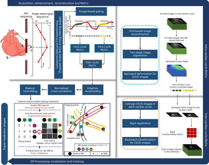
Myocardial microvasculature and haemodynamics are indicative of potential microvascular diseases for patients with symptoms of coronary heart disease in the absence of obstructive coronary arteries. However, imaging microvascular structure and flow within the myocardium is challenging owing to the small size of the vessels and the constant movement of the patient’s heart. Here we show the feasibility of transthoracic ultrasound localization microscopy for imaging myocardial microvasculature and haemodynamics in explanted pig hearts and in patients in vivo. Through a customized data-acquisition and processing pipeline with a cardiac phased-array probe, we leveraged motion correction and tracking to reconstruct the dynamics of microcirculation. For four patients, two of whom had impaired myocardial function, we obtained super-resolution images of myocardial vascular structure and flow using data acquired within a breath hold. Myocardial ultrasound localization microscopy may facilitate the understanding of myocardial microcirculation and the management of patients with cardiac microvascular diseases.


